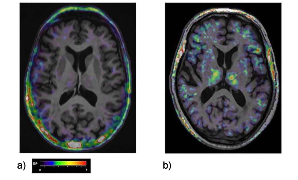
[11C](R)-PK-11195 PET BP maps co-registered to the individual MRI scan of a) a healthy control, b) of a MCI patient. BP increases can be seen fronto-temporal areas as well as in the thalamus. The color bar denotes BP values from 0 to 1

[11C](R)-PK-11195 PET BP maps co-registered to the individual MRI scan of a) a healthy control, b) of a MCI patient. BP increases can be seen fronto-temporal areas as well as in the thalamus. The color bar denotes BP values from 0 to 1
The authors thank study personnel at the Memory Assessment and Research Centre, Southampton, the Institute of Brain, Behaviour and Mental Health, and the Wolfson Molecular Imaging Centre, University of Manchester who were involved in administration, drug delivery, venesection, sample preparation, and imaging procedures. The authors also thank all patients and carers who took part in the study.
AG, AHJ, KH and CH: Conceptualization, Funding acquisition. AG, CH, RS, TG, KM, EV and IL: Investigation, Data curation, Formal Analysis. CH and FT: Formal Analysis. AG and CH: Writing—original draft. All authors: Writing—review & editing.
The authors report no conflict of interest.
The protocol and consent forms were approved by a multi-centre research ethics committee (South Central-Hampshire A Research Ethics Committee), reference number 15/SC/0435. The study was performed in accordance with the Declaration of Helsinki and principles of Good Practice. An independent data and safety monitoring board monitored adverse events.
Informed consent to participate in the study was obtained from all participants.
Not applicable.
The data that support the findings of this study are available from the corresponding author upon reasonable request.
The study was funded by the Alzheimer’s Society UK, Alzheimer’s Drug Discovery Foundation and the European Union Seventh Framework Programme [FP7/2007-13] INMIND. Grant agreement number 278850. The funders had no role in study design, data collection and analysis, decision to publish, or preparation of the manuscript.
© The Author(s) 2023.