Affiliation:
1Centre for Nanosciences, University of Okara, Okara 56130, Pakistan
ORCID: https://orcid.org/0009-0000-4701-0503
Affiliation:
1Centre for Nanosciences, University of Okara, Okara 56130, Pakistan
ORCID: https://orcid.org/0009-0007-9969-9630
Affiliation:
2Department of Chemistry, University of Picardie, 80000 Amines, France
ORCID: https://orcid.org/0009-0002-6345-3894
Affiliation:
3Department of Zoology, Faculty of Life Sciences, University of Okara, Okara 56130, Pakistan
ORCID: https://orcid.org/0000-0003-2974-4823
Affiliation:
3Department of Zoology, Faculty of Life Sciences, University of Okara, Okara 56130, Pakistan
ORCID: https://orcid.org/0009-0002-0586-9066
Affiliation:
1Centre for Nanosciences, University of Okara, Okara 56130, Pakistan
ORCID: https://orcid.org/0000-0002-3474-7458
Affiliation:
1Centre for Nanosciences, University of Okara, Okara 56130, Pakistan
ORCID: https://orcid.org/0000-0002-5166-3328
Affiliation:
1Centre for Nanosciences, University of Okara, Okara 56130, Pakistan
ORCID: https://orcid.org/0000-0002-1240-7401
Affiliation:
1Centre for Nanosciences, University of Okara, Okara 56130, Pakistan
ORCID: https://orcid.org/0000-0002-7338-2834
Affiliation:
1Centre for Nanosciences, University of Okara, Okara 56130, Pakistan
Email: misbahullahkhan143@uo.edu.pk
ORCID: https://orcid.org/0000-0003-2960-103X
Explor BioMat-X. 2024;1:366–379 DOI: https://doi.org/10.37349/ebmx.2024.00025
Received: September 29, 2024 Accepted: December 06, 2024 Published: December 13, 2024
Academic Editor: Omid Akhavan, Sharif University of Technology, Iran
The article belongs to the special issue Plasmonic Nanostructures for Designing Optical Biosensors
Silver iodide (AgI) nanostructures have been considered as promising candidates for optical biosensors owing to their optical characteristics of optical properties, including tunable surface plasmon resonance (SPR) and fluorescence enhancement. Such properties let one analyze biomolecules with high sensitivity, which makes them ultra-useful in diagnostics. The formed AgI nanostructures can be synthesized using chemical precipitation and template methods that enable fine-tuning of the morphology and crystallinity of the final nanostructure. The presence of SPR enhances optical signals potentially, and fluorescence enhancement helps visualize biomolecule interactions easier as the analyte concentration is usually low. Such uses of biosensors include applications in proteins, nucleic acids, and other biomolecules for progress in disease diagnosis and pharmacogenomics. Moreover, the good biocompatibility level of the created AgI nanostructures makes it possible to integrate them into biological systems safely, increasing their usage in medicine. This integration of their appealing optics, biosensing operating principles, and biocompatibility establishes their centrality in the creation of future photonic biosensors for faster, intuitive, and painless detection.
Due to the high selectivity and sensitivity of optical biosensors for the analysis of biological molecules, the amount of research related to these devices has increased significantly in the past several years. It has been clearly demonstrated that the method of speciation analysis and its application as an instrument for evaluating the threat to human health or for establishing the quality of food products are of great importance [1]. Optical biosensors have several qualities that are especially desirable in contrast with other normal analytical methods. These advantages include the capability to target a large number of biological and chemical species with direct and immediate analyses without the necessity of tags. These devices offer additional advantages due to their high specificity and sensitivity, compact design, and relatively low cost. Microelectronics, microelectromechanical systems (MEMS), micro/nanotechnologies, molecular biology, biotechnology, and molecular chemistry are among the numerous technologically sophisticated ideas used in the process of creating new optical biosensors [2]. The presented review forms a constructively ordered scientific database of research and technological development concerning optical biosensors over the past decade. A particular increase is observed in the use of biosensors, specifically in healthcare applications, environmental monitoring, and the field of biotechnology [3].
From the point of view of the mode of signal transduction (optical, electrochemical, thermometric, piezoelectric, and magnetic), it is possible to divide biosensors into a large number of subtypes. The biosensors that are widely recognized in the media and are regarded as optical biosensors are the type of biosensors that get the highest attention. The optical detection process is achieved by harnessing the interaction between the light field and a biorecognition component [4]. It is possible to employ two basic categories for distinguishing optical biosensing technologies. In this system, these categories are label-free modes and label-based systems. When the mode is label-free, the detected signal has been produced purely from the interaction of the substance being studied with the transducer. In layman’s terms, what this state is that the signal was generated directly. On the other hand, label-based sensing was characterized by the presence of a label when the optical signal is reconstructed through the colorimetric, fluorescent, or luminescent method. About label-based sensing, the level of distinguishing a label was characterized according to the type of label [5]. As an example, the comparatively simple sugar, glucose, may be detected by employing enzymatic oxidation in combination with label-assisted sensing for identification. Glucose analysis in blood has been the most commercially successful application of a biosensor, up to this point. The handheld glucose meter, widely utilized by individuals with diabetes for continuous monitoring, represents a notable economic success among biosensors. Its widespread adoption and practicality underscore its commercial viability and effectiveness in the biosensor market. Labeling, in contrast, may change the binding properties, thus leading to the introduction of systematic inaccuracy into the biosensor analysis. Specifically, when one of the bioactive chemicals has an attached label, such as the interaction between an antibody and an antigen, this is the case [6].
Imposing a biorecognition sensing element, one of the components of an optical biosensor is a miniature analytical instrument (Figure 1) attached to an optical transducer system. A key goal for the design of an optical biosensor is to create a signal that is proportional to the concentration of a substance (analyte) that is measured. This signal is produced by the optical biosensor we are discussing. The optical biosensor and the receptor alone are used to create this signal. Depending on the nature of the target analyte, it is possible to use various types of biological molecules with the optical biosensor. Such molecules include enzymes, antibodies, antigens, receptors, nucleic acids, whole cells, and tissues. In such cases, it is the biorecognition element that is primarily responsible [7]. Three methods take advantage of the evanescent field, which is formed just above the surface of the biosensor. Among them, there are three types: surface plasmon resonance (SPR), evanescent wave fluorescence, and optical waveguide interferometry. Other methods include optical waveguide interferometry. Such methods are used in a systematic manner to discover the event that happens between the biorecognition component and the analyte. Even though the construction of optical biosensors can be described by a large number of variations, the purpose of this review is to focus on several of them. The choice of these variants was due to their versatility and their propensity to identify compounds critical in biological processes. It has therefore been achieved to incorporate nanomaterials, especially nanostructures of silver iodide (AgI), into optical biosensors [8].
Long et al. [9], in their study in 2013, have shown that this has led to performance enhancement of these devices for applications in disease diagnosis, air and water quality analysis, and food quality. A great number of fields have adopted applications for the use of optical biosensors. Reduced AgI nanostructures are ideal for increasing the sensitivity and selectivity of biosensors because of their extraordinary optical properties, namely bright luminescence, tunable light absorption, and pronounced SPR effects [10]. These properties make AgI nanostructures potential candidates for this purpose, as they exhibit these features in their behaviors. Consequently, while all these properties have been taken into consideration, AgI nanostructures are simply wonderful options. As a result of these characteristics, it is possible to translate biological signals into detectable optical outputs in a sufficiently efficient manner to enable the identification of target analytes in relatively short times once the conversion has occurred. Of the various elements that comprise these analytes, there are pathogens, proteins, enzymes, and varieties of nucleic acids [11].
AgI nanostructures are biocompatible and have accessible surface chemistry that allows the accretion of certain proteins, thereby increasing the selectivity of the optical biosensors. In addition, some proteins can adsorb onto AgI nanostructures. One strength is that this is true to a large extent. Innovative architectures of AgI nanostructures have been developed with a high level of control over the features of the engineered material, such as size, shape, and distribution of nanopatterns on fabric. This was not possible before. This has made it possible to have a more refined management over a number of aspects of the problem. Innovations have made it possible for the creation of resonant systems that can be used for biosensing and preferably be designed to respond based on user features [12]. Thus, the techniques that have improved the detection limits and reaction times have made the optical biosensors more useful for practical applications. This has been due to their enhanced means of measurement. Moving from medical diagnosis only, the applications of AgI-based biosensors are expanding. Moreover, those biosensors are used to check environmental pollutants and toxins and food safety by detecting contaminants present in the food chain. These applications ensure food safety, meeting the objectives of the project. They are especially useful tactical tools because they may be used across a range of industries [13].
AgI is understood to be a material with excellent optical characteristics, including high refractive indexes and ratios between light and matter energy and other materials, according to [14]. AgI is a chemical compound that is well known for having great optical properties. With a great superionic conductivity higher than 1 Ω−1 cm², AgI is widely known as an effective ionic conductor. This conductivity was realized in the α-phase of this chemical compound when it was subjected to high temperatures. Highly disordered phase structure is the cause of the ionic conductivity that is observed inside this material [15]. From the observation, it has been demonstrated that as the temperature gets below 146°C, it causes a phase transition of AgI to α-AgI, which is known as a super ionic conductor [16]. At room temperature, it has a blatant band gap of 2.9 eV, which is the reference point at which the temperature of the material is normally measured. Concerning the particle size dependence of the substance, the ionic conductivity and the phase transition temperature appeared to be sharply affected. AgI is widely used in a number of industrial segments such as photography, medical, antibacterial, electrical, magnetic, and optical [17]. With regard to the synthesis of AgI nanoparticles (NPs), there are a number of methods and techniques that have been employed. These techniques include ultrasonic, mechanochemical, hydrothermal, and laser ablation of silver in a solution containing sodium dodecylsulfate surfactant and iodine, reverse micelles methods, electrochemical, and ultrasonic spray pyrolysis [18].
Different nanostructures of AgI and their uses are depicted in the following Figure 2. AgI nanostructures, including NPs, nanorods, and nanospheres, possess different optical, catalytic, and antibacterial characters. Having these features makes them very effective in several applications, including medicinal uses, wound dressing, anti-microbial activities, environmental applications, and as catalysts in chemical reactions. Besides, the tunability of AgI nanostructures is even more beneficial in different industrial and biomedical applications.
Meaningfully, nanostructures made out of AgI possess many plasmonic properties, allowing for more effective control of these properties at the nanoscale. This is due to the small size of the nanoscale. In light of this, it is also possible to perform further refinements to these characteristics. These nanostructures can be described as attractive prospects for the photonics sphere, which essentially means that they can be scrutinized. Because this is the case, they have the capacity for recognition on this basis [19].
Chemical vapor deposition (CVD) techniques, sol-gel techniques, and electrochemical techniques have been established for synthesizing nanostructures of AgI [20]. These systems have been applied in the synthesis of nanostructures. This represents a brief description of the techniques that can be used to synthesize AgI nanostructures [21]. The below Figure 3, represents different approaches to synthesizing of AgI nanostructures.
Nanostructures or thin films of AgI can be formed by a chemical deposition technique called CVD, where gaseous precursors undergo a reaction with a preheated substrate. By applying this technique, it is possible to gain improved control over the size and size distribution of the nanostructures, resulting in the synthesis of highly pure and uniform AgI nanostructures [22].
The CVD method for preparation of AgI is further depicted in the schematic diagram in Figure 4. Here, the metallic silver precursor vapor is deposited on iodine gas and transformed into AgI nanostructures in a reaction chamber at high temperature. The method enables manipulation of the shape and size of the synthesized NPs, hence suitable for forming high-quality AgI for numerous purposes, including catalysis and as antimicrobial agents. The CVD technique is well preferred for yielding high-purity, uniform, and easily scalable AgI nanomaterials.
The sol-gel method involves the transition of a liquid-phase sol into a solid-gel phase. This transition is fundamental to the development of the technique. To synthesize AgI metal nanostructures, silver sources are mixed with iodine precursors before the fabrication of the nanostructures. Once the gelation process is complete, the material undergoes further drying and annealing steps [23]. In summary, this process results in densely packed nanostructures with control over particle size. Additionally, AgI nanostructures can be created at low temperatures, which is a notable advantage [24].
Figure 5 describes a schematic of the sol-gel method for synthesizing AgI. In this process, silver salts are kept in a solvent and converted into sol, and then the sol is mixed with an iodine precursor. Sol converts into a gel, and the gel further solidifies into a three-dimensional network, and finally, on heating, AgI nanostructures are formed. This permits the synthesis of highly surface area and property uniform AgI under controlled conditions.
Electrochemical synthesis is performed by passing an electric current through a solution containing silver and iodide ions to initiate various processes. This technique is employed to produce the desired chemical compound and results in the formation of AgI nanostructures on the electrode surface [25]. This procedure allows for precise control over the composition and shape of the nanostructures by adjusting the voltage and other reaction conditions. Both composition and shape depend on these controlled properties to achieve the desired specific structure [26].
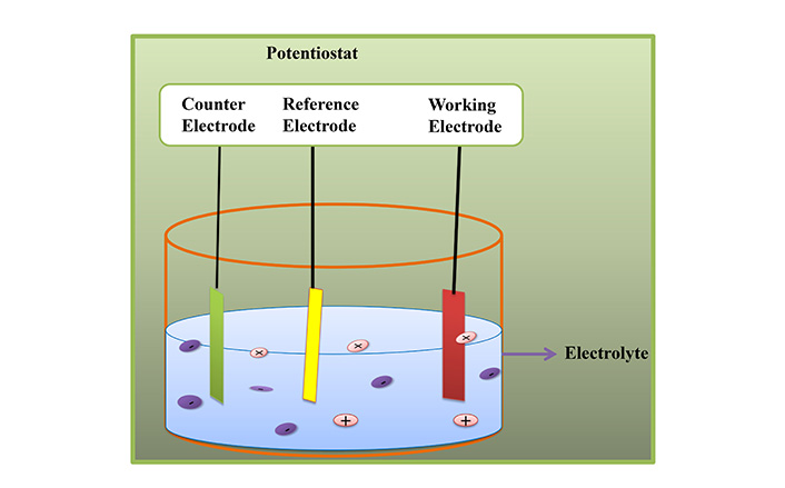
The electrochemical method for the preparation of AgI is given in Figure 6. During this process, two silver electrodes are mounted in an iodide ion solution, which is an electrolyte solution of some sort. An electric current is passed through the solution where, at the cathode, the silver ions are reduced on the surface of the electrode and then react with iodide ions to produce AgI nanostructures. This method can provide excellent control of aspects such as particle size and shape, which is beneficial in sensor, catalytic, and antimicrobial applications.

Representation of schematical view of electrochemical method for the preparation of AgI
These techniques permit precise regulation of the dimensions, morphology, and crystalline configuration of AgI nanostructures, which in turn affects their optical and sensing characteristics [27].
The efficiency of biosensors based on AgI nanostructures relies primarily on features such as SPR and enhanced scattering. SPR is defined by the oscillation of free electrons, a process that occurs in AgI nanostructures when they are illuminated. This increases the intensity of the surrounding electromagnetic field manifold, especially with environmental changes like refractive index, as shown in Figure 7. Specific proteins and DNA can bind to these nanostructures, altering their refractive index. Such changes result in variations in the SPR signal, allowing real-time analysis of biomolecular interactions without the need for labels. These nanostructured materials, such as AgI, have potential applications in disease diagnostics, environmental monitoring, and quality control, including testing for food safety regulations [28].
Some of the optical features of AgI nanostructures are shown in Figure 7. These properties comprise the absorption, reflection, and scattering effects, and all these are highly dependent on the size, shape, or crystallinity of the nanomaterial involved. This property also affords AgI unique light adsorption in the UV-visible region and light scattering that is essential in optoelectronics, sensors, and photocatalytic applications. Due to tunable optical characteristics, AgI can be used in many unconventional applications of modern technologies.
Increase in scattering is another important optical property of AgI nanostructures; increasing this can lead to an enhancement in the observed signal. This characteristic is highly desirable in the biosensor’s domain, as biosensors often require detection of low concentrations of an analyte. It holds significant value in this field [29]. The enhanced ability of AgI nanostructures to induce light scattering makes the sensor more sensitive and lowers its limit of detection. This structure is achieved by using AgI nanostructures to improve light dispersion. By controlling the size, shape, and structural properties of AgI nanostructures, researchers can modify the optical properties and tailor them for biosensing applications [30]. As a result, AgI nanostructures have evolved into versatile and valuable components in the development of next-generation diagnostic devices [31].
As a result of the target analyte, most optical biosensors must undergo several functional steps, often including absorption, emission, scattering, and similar processes. The interactions observed between the sensor surface and biomolecules cannot be explained without the signals produced from these inputs. Ong and co-authors [32] demonstrated in their 2021 study that optical detection can be made more sensitive and accurate by using AgI nanostructures as photonic amplifiers. The optimization of SPR and sensitivity to the refractive index allows the AgI nanostructures to enhance the interaction of the analyte with light. The authors noted that signal generation and detection capabilities remain enhanced, even in low analyte concentration situations. Several advantages of integrating AgI nanostructures as photonic amplifiers in biosensors include improved efficiency, enhanced scattering, and absorption processes [33].
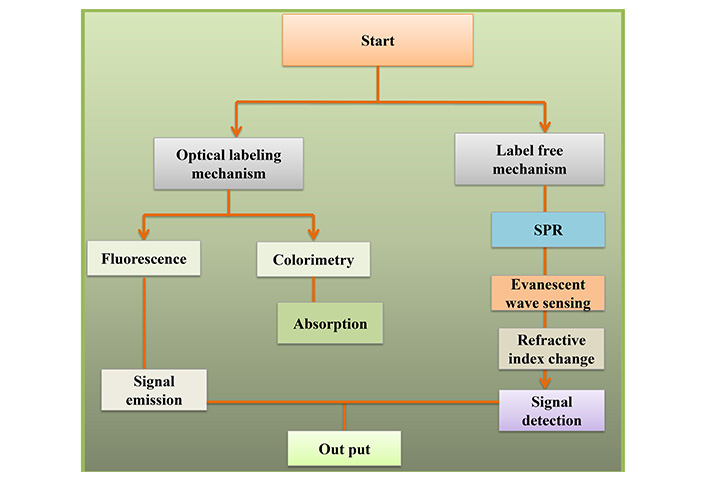
The schematic mechanism of an optical biosensor is illustrated in Figure 8. In this process, there is an attempt to attach a biological recognition element, which includes enzymes/antibodies, to a substrate and interact with the target analyte. The interaction between the optical sensor and the material under test alters one or more properties of light, for instance, reflection, absorption, or fluorescence. They are then quantified, and the data are compared to check for the existence of the analyte and the intensity of the same, making it very efficient in diagnostics and in real-time monitoring.

Representation of schematical mechanism of optical biosensor. SPR: surface plasmon resonance
Enhanced signal resolution and clarity due to increased scattering have also been achieved, enabling the observation of minute optical response changes in the analyte solution. The intensification of dispersion makes this possible. Based on these optical properties, the size, shape, and crystallinity of AgI nanostructures can be designed to achieve specific aspects of biosensing functionality [34]. For this reason, they can be tailored to better fulfill these requirements, making them suitable for environmental monitoring and other sensitive medical applications. There is potential for real-time enhancement of detection and monitoring of biological and chemical agents using optical biosensors constructed with AgI nanostructures [35].
Among the applications of AgI nanostructures, the biosensing technique called SPR is probably the most commonly used. Nevertheless, this application is among the most profitable of all of them. Lopes et al. [36] stated that localized surface plasmons in AgI nanostructures are activated through incident light. This activation yields strong optical signals that can be possibly followed for biosensing purposes. These transformations occur in these processes whenever biomolecules, including proteins or DNA, interact with the surface of the sensor [37].
Additionally, AgI nanostructures possess the potential to enhance fluorescence signals through a method known as metal-enhanced fluorescence (MEF). Another technique mentioned is the amplification of fluorescence signals, which increases the emission intensity when fluorophores are located near AgI nanostructures [38]. This effect greatly assists in the development of fluorescence-based biosensors, enabling the detection of analytes at trace levels, as described by [39]. These authors further noted that this effect is particularly beneficial in this process.
AgI nanostructures are used for opto-electrical biosensing applications due to their high sensitivity. AgI nanostructures are employed in diagnostic procedures by identifying DNA, biomarkers, and disease-associated proteins at very low concentrations. Early detection of diseases such as cancer and infections, for instance, increases the patient’s chances of survival. Biosensors made from AgI enable quick diagnosis because of the high sensitivity and selectivity of the compound [40]. Pollutants remain a major concern to human life because they impact the quality of soil, water, and air. One significant advantage of these biosensors is their ability to identify organic contaminants and trace heavy metal pollution that contributes to environmental degradation. The AgI nanostructures enhanced optical properties facilitate a strong environmental response, allowing the detection of even minute pollutants in the environment [41].
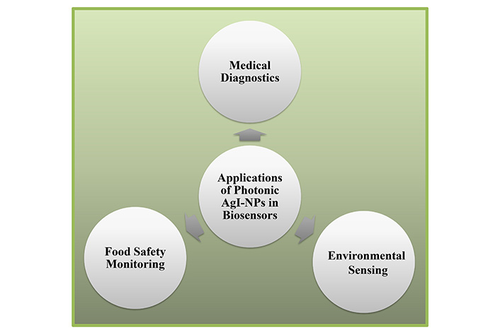
As highlighted in Figure 9, a number of applications of AgI-NPs have been pointed out in environmental, food safety, and medical aspects. Within the environmental area, a vast application of AgI-NPs is seen in water treatment and in the degradation of pollutants. In food safety, they act as preservative agents to curb microbial load in foods to be preserved. In medical applications, AgI-NPs are used in wound dressing and drug delivery systems because AgI-NPs have bactericidal properties and biocompatibility. It is due to their versatile applications that make AgI-NPs more applicable in various sectors of the economy.

Representation of schematical view of application of AgI-NPs. AgI-NPs: silver iodide nanoparticles
They help to filter out toxins, bacteria, and viruses in foods so that people can consume safer food products. Such nanostructures have the potential to prevent foodborne contaminants from harming people by identifying them in foods before reaching the market. Real-time and accurate detection of pollutants across industries to safeguard public health is made possible by biosensors based on AgI nanostructures [42].
Newly developed accurate diagnostic instruments can identify bio-signatures linked to diseases such as cancer and other illnesses, including those generated in the medical industry with nanostructures of AgI [43]. These devices enable the detection of biomarkers related to diseases. This development in medical devices has led to significant improvements in early disease identification. Due to this progress, there have been large successes in completing these diagnostic advancements [44].
Other types of optical biosensors detect pollutants in air and water by using AgI nanostructures. These biosensors enhance the process of monitoring environmental pollutants to an unparalleled degree. Li et al. [45] highlighted such sensors’ effectiveness in differentiating toxins. Additionally, these contaminants include microbes that cause illness if ingested, as well as heavy metals and pesticides [46].
Foodborne communicable diseases and pollutants pose major threats to public health. Biosensors incorporating AgI can detect potentially harmful microbes, toxins, and chemical contaminants in food due to advancements in optical biosensors [47]. This capability greatly enhances food safety standards, which was once thought nearly impossible [48]. This breakthrough, achieved by Zhang and colleagues [49] through rigorous investigation, helps ensure and verify food industry quality and safety.
Despite the opportunities offered by AgI nanostructures in optical biosensing, some challenges are associated with their usage. These challenges include issues of toxicity, long-term stability, and cost, as discussed. Addressing these challenges is essential for the successful commercialization of AgI-based biosensors, as they present potential obstacles to widespread adoption [50].
One well-known issue is the potential toxicity of silver-based NPs, especially in food and medical applications. Developing new biocompatible coatings or formulations that reduce toxicity while preserving optical properties is a priority [16]. These coatings must be compatible with various organisms.
AgI nanostructures are prone to degradation under harsh environmental conditions, particularly at high temperatures. This degradation is more significant in nanostructures due to their small size. Researchers are exploring protective coatings and surface treatments to increase stability and facilitate large-scale manufacturing [51]. Large-scale production is essential for making these nanostructures cost-effective [52].
Advancements in photo-catalytic AgI nanostructures and photonic AgI nanostructures hold promise for developing biosensors that can detect multiple analytes with high sensitivity. These developments represent the future of biosensing research, with new NPs, nanofabrication techniques, and material sciences contributing to the next generation of optical biosensors [53].
Incorporating AgI nanostructures with other materials, such as graphene or quantum dots, could lead to advanced hybrid biosensors with higher sensitivity, selectivity, and multiplexing capacity [54]. A study by Nawito [55] demonstrated that hybrid systems outperformed conventional analysis systems by at least an order of magnitude in these aspects.
Current research is focused on developing smart biosensors that integrate artificial intelligence (AI) and machine learning (ML) for real-time data processing and analysis. Bhaskar et al. [37] suggest that these systems, with their rapid and accurate diagnostic capabilities, have the potential to transform medical and environmental surveillance by providing quick and reliable diagnoses. This capability enhances biosensing applications across various sectors, allowing these devices to deliver precise diagnostic outcomes efficiently [39].
Nanostructures are fairly new in the field of optical biosensing, and those made of AgI have been rated as very efficient instruments. These nanostructures have proved to be strong tools because of the tremendous improvement in sensitivity and selectivity due to their use. Thus, there is a great deal of potential for using biosensors based on AgI, even though there are issues, such as toxicity and stability, on the horizon. It might be that this promise will be realized in the future through further research and development. They are considered more important today in sensing technologies due to the increasing role they play in diverse fields, including disease detection, environmental assessment, quality of foods, and others.
AgI: silver iodide
CVD: chemical vapor deposition
NPs: nanoparticles
SPR: surface plasmon resonance
HA: Writing—review & editing. NN, A Ullah, and MWA: Visualization. MM, MJS, and MA: Formal analysis. A Umar: Data curation. SH: Resources, Investigation. MUK: Conceptualization, Investigation, Methodology, Project administration, Resources, Software, Supervision, Validation.
Not applicable.
Not applicable.
Not applicable.
Not applicable.
Not applicable.
Not applicable.
© The Author(s) 2024.
Copyright: © The Author(s) 2024. This is an Open Access article licensed under a Creative Commons Attribution 4.0 International License (https://creativecommons.org/licenses/by/4.0/), which permits unrestricted use, sharing, adaptation, distribution and reproduction in any medium or format, for any purpose, even commercially, as long as you give appropriate credit to the original author(s) and the source, provide a link to the Creative Commons license, and indicate if changes were made.
Hamid Nadjari, Hadi Movahedinejad
