Affiliation:
Robotics Laboratory, School of Computer Science and the Environment, Liverpool Hope University, L16 9JD Liverpool, UK
ORCID: https://orcid.org/0000-0002-3250-8066
Affiliation:
Robotics Laboratory, School of Computer Science and the Environment, Liverpool Hope University, L16 9JD Liverpool, UK
ORCID: https://orcid.org/0000-0002-8157-5003
Affiliation:
Robotics Laboratory, School of Computer Science and the Environment, Liverpool Hope University, L16 9JD Liverpool, UK
Email: seccoe@hope.ac.uk
ORCID: https://orcid.org/0000-0002-3269-6749
Explor Digit Health Technol. 2024;2:346–359 DOI: https://doi.org/10.37349/edht.2024.00033
Received: July 18, 2024 Accepted: November 07, 2024 Published: November 25, 2024
Academic Editor: Shariful Islam, Deakin University, Australia
The article belongs to the special issue Wearable Technologies and Application of Machine Learning in Healthcare
This research provided an in-depth analysis of endoscopic capsules as an innovative application of the Internet of Things (IoT) in healthcare. The study revealed the importance of these systems in advancing gastrointestinal diagnostics due to their non-invasive nature and ability to provide comprehensive internal imaging. The work systematically investigated the device’s technical design, power management strategies, communication protocols, and how it performs its secure and efficient operations. Findings from this analysis highlighted the transformative impact of these capsules despite current constraints, such as battery limitations and procedural costs. Ultimately, this wide review confirmed that endoscopic capsules redefine medical diagnostics, fusing patient comfort with innovative technology. Moreover, as developments continue, these devices have promising potential to shape the future of intelligent, interconnected healthcare solutions.
The ongoing Fourth Industrial Revolution, which was initiated in the late 1990s, marked a significant technological leap which has been driven by rapid progress associated with the global adoption of the internet technology and significant advancements in computer science [1]. At the heart of this revolution is what is called Internet of Things (IoT), namely a set of networks built upon the generation of big data through the growth in connectivity and information-capturing devices [2]. The IoT functionality orbits around a vast diversity of embedded sensors, specialized data processing systems, and algorithms. According to the continuous spread of these devices, the IoT network succeeds in offering an assistive personalized awareness and progressive flow in data analysis across various fields.
Whether operating individually or clustered, these IoT-connected devices extend the capacity of centralized computational systems, such as cloud computing or data centers, by means of bridging the processing power directly to the data source. With a global tendency to fusion physical and digital technologies, IoT has become the key innovative industry, which is crucial for advancing systems that demand extensive amounts of data, such as artificial intelligence (AI), robotics, genetic engineering, and quantum computing.
The goal of the IoT network is to provide a deeply interconnected world where devices can communicate with each other in order to automate processes and improve efficiency, sustainability, and economic benefit [3].
The versatility of the IoT technology is vividly illustrated through a simple analysis of the worldwide number of IoT connections over the last few years: according, for example, to the ISE 2022 statista.com website (Transforma Insights), the number of consumer IoT connections in 2018 was 3.14 billion, whereas in 2028 it is foreseen to have 20.30 billion of connections. Similarly, smart cities’ IoT connections would rise from less than 0.19 billion in 2018 to 5.29 billion in 2028. The IoT market distribution 2024 and projections overview predicts a revenue by the segment of 2,226 billion US dollars (USD) in 2028 vs. an overall revenue of less than 471 billion USD in 2018 with more than 30% of the IoT market devoted to the healthcare sector.
In the context of daily life, within a smart home the IoT devices enhance comfort and energy efficiency through automated control of household functionalities [4]. Similarly, an urban infrastructure can become more intelligent through the use of IoT technology for traffic management and energy conservation, providing more sustainable solutions and communities [5]. In the agricultural field, using sensors and data analytics enables precise farming methods, optimization of the resources, and better yields [6]. The industrial sector benefits from its own specialized network of IoT-connected systems as well, since they can improve process optimization and supply chain management, with a crucial role in economic advancements and operational efficiencies (Figure 1) [7].

Timeline of ingestible capsule technologies. AI: artificial intelligence
Note. Adapted from [8], CC BY 4.0.
One of the most significant impacts of IoT technologies is in healthcare, where innovations can affect and improve the level of patient care and the diagnostic accuracy. The IoT devices enhance health data collection, provide valuable insights into the human body, and enable the remote monitoring of patients [9]. These capabilities mark a shift towards more efficient and personalized diagnostics, directly addressing some of the long-standing challenges associated with medical procedures, such as traditional endoscopy. While indispensable for diagnosing gastrointestinal (GI) disorders, traditional endoscopic procedures have limitations, including restricted visualization, uncomfortable preparations, sedation requirements, and procedural risks. Moreover, their capability to thoroughly examine the small intestine (a vital section of the GI tract) is limited [10]. Endoscopic capsules (ECs) have emerged as a breakthrough solution, offering a minimally invasive and patient-friendly method to examine the entire digestive tract.
The concept of an ingestible device for diagnostic imaging was conceived in the early 1950s [11], but technological constraints delayed its practical development for decades. Advancements in microelectronics, optics, and wireless transmission in the late 1990s made capsule endoscopy a viable innovation [12]. Once approved by the U.S. Food and Drug Administration (FDA) in 2001, these devices began to incorporate the principles of IoT, redefining medical diagnostics.
By leveraging advanced sensors, wireless data transmission, and miniaturized electronics, EC systems connect to the internet, enabling the continuous transmission of collected data to the cloud. These characteristics allow the information to be accessible through centralized data systems, enabling medical practitioners to monitor, analyze, and store information in real-time. Such a patient-specific closed-loop IoT network provides immediate access to the endoscopic diagnostic data and integrates it with the person’s health records. This integration enhances patient care, offering a fast and more holistic view of the individual’s health status, thereby revolutionizing the process of GI diagnostics and patient management [8].
These peristaltic-travelling capsules provide a comprehensive visualization of the GI tract, previously unattainable, bringing ease and comfort to patients and offering valuable insights to medical practitioners [13, 14].
This review paper explored the technical aspects of capsule endoscopy as an IoT device, examining its components, power management, communication protocols, security considerations, and deployment challenges. By investigating these aspects, the research aimed to provide a comprehensive understanding of this innovative technology and its potential to improve and further advance GI diagnostics.
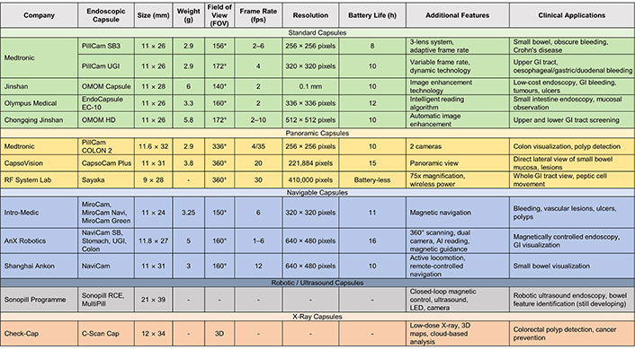
In modern medical technology, the EC represents a significant advancement in gastroenterology and broader healthcare applications (Figure 2). It is a novel, non-invasive, and autonomous method for visually examining the GI tract. This device, encased in a biocompatible, non-digestible shell of polyethylene or polymethyl methacrylate (PMMA), measures 11 mm in diameter and 25–31 mm in length and weighs between 2.9 and 4 g (Figure 3, left panel) [14]. Its smooth design and pill-like dimensions facilitate easy ingestion and ensure safe transit through the digestive system via natural peristalsis. During this transit, the capsule continuously captures high-resolution images transmitted to an external receiver. The data received provides critical insights into areas traditionally difficult to reach with conventional endoscopy, enabling physicians to diagnose and monitor various GI disorders remotely.

An overview of current ECs in the market. AI: artificial intelligence; ECs: endoscopic capsules; GI: gastrointestinal; LED: light-emitting diode
The typical EC consists of several key components engineered together to provide a comprehensive visualization of the GI system.
ECs use up to four CMOS (complementary metal-oxide-semiconductor), cameras chosen for their low power consumption, compact size, and high-speed imaging capabilities (Figure 3, right panel). Unlike their charge-coupled device (CCD) counterparts, CMOS sensors process the charges into digital signals at the sensor’s location. This direct conversion process reduces power consumption and allows for faster processing speeds. Each pixel in a CMOS sensor has its own charge-to-voltage conversion and includes amplifiers, noise-correction, and digitization circuits, making the CMOS technology highly efficient.
The cameras are fitted with wide-angle and short focal lenses, providing a field of view (FOV) ranging from 140° to 360°, depending on the specific device. Additionally, arrays of white light-emitting diodes (LEDs) are strategically placed around the cameras to evenly illuminate the environment. This overcomes one of the CMOS low-light performance limitations and ensures optimal image quality even in the darkest regions of the digestive system.
Furthermore, the use of CMOS technology addresses the need for energy efficiency, allowing for longer operational time critical for medical applications, while the capsule’s steady progression minimizes the impact of the rolling shutter effect, a well-known disadvantage in these optical sensors during image capture. These image systems typically offer a resolution of 256 × 256 to 640 × 480 pixels, with a variable sampling rate adapted to their hardware optimization performance, which can be 2–35 frames per second (fps), being able to provide a detailed visual map of the esophagus, stomach, and intestines. This implementation is a classic engineering example of leveraging the best of current technology to adapt and improve a device.
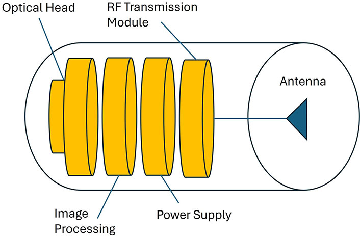
The application-specific integrated circuit (ASIC) forms the core of the image processing system within an EC. This specialized semiconductor efficiently performs several critical functions. Primarily, the ASIC handles real-time image processing, including initial capture, digital conversion, and image compression. Its design focuses on managing the vast amount of data generated by the CMOS cameras and compressing it into a format that balances image quality and file size for effective wireless transmission (Figure 4) [14]. This process is crucial for maintaining a high-quality diagnostics image while constrained by the capsule’s limited power resources.

Wireless medical capsule endoscopy system diagram. Images from the optical head are locally processed and feed the RF transmission module providing wireless communication through the antenna. RF: radio-frequency
The ASIC also has a critical role in power management. It optimizes the image processing workflow to reduce energy consumption, extending the capsule’s battery life. Innovative circuit design minimizes idle power usage and maximizes the efficiency of active processing tasks, achieving optimal operational state.
ECs rely on a radio-frequency (RF) wireless system to transmit data to an external receiver (uplink), typically operating within the 400–440 MHz ultra-high frequency (UHF) band. This frequency range is chosen for its ability to quickly penetrate the human body with minimal signal loss, ensuring that high-resolution images captured by the capsule are transmitted efficiently to the external receiver, with a maximal transmission distance of up to 3 m (Figure 4).
RF wireless transmission sends data wirelessly using radio waves, converting digital data into electromagnetic signals that can travel through various mediums, including air and biological tissues. The RF transmitter within the capsule encodes the captured images onto these radio waves, which get received by an external device, further demodulated, and re-converted into digital data.
The design and operation of the capsule’s RF transmission system are meticulously engineered to maintain a stable communication link with the external receiver. This aspect ensures the reliable transmission of all captured images. The integration of RF transmission technology within the capsule is justified by its proven reliability and safety for use within the human body. The transmission power, carefully regulated to prevent potential harm or discomfort to the patient, adheres to stringent medical and communications regulations, including Federal Communications Commission (FCC) guidelines [13], International Electrotechnical Commission (IEC) standards for electromagnetic compatibility, and European Telecommunications Standards Institute (ETSI) standards in Europe for health and safety under the radio equipment directive [14].
Additionally, the UHF telemetry system’s design is specifically tailored to address the unique challenges of transmitting data through biological tissues. It optimizes signal strength and modulation techniques to ensure consistent performance and overcome potential issues such as signal attenuation or interference.
The external wearable receiver, featuring advanced RF receiving technology, captures and stores the transmitted data. It is equipped to demodulate the signal from the capsule and reconstruct the compressed images (usually jpeg for images or mpeg for videos), making them available for medical professionals to review [15].
This sophisticated integration of ASIC processing and RF wireless transmission technology forms the backbone of the functionality of ECs. It is a remarkable example of engineering innovation, combining detailed visualizations of the GI tract with the technological fusion of IoT capabilities. This aspect facilitates the diagnosis and monitoring of conditions with unprecedented ease and efficiency, showcasing the potential of modern medical devices to improve patient care.
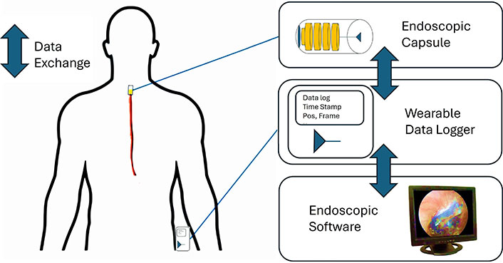
ECs integrate advanced communication protocols to ensure the accurate and secure transmission of diagnostic data from the capsule to the external receiver and further to the cloud or its dedicated workstation (Figure 5). These protocols encompass a set of technical standards and procedures that dictate data encoding, transmission, and decoding, safeguarding its integrity and confidentiality.

The endoscopic capsule is swallowed into the digestive system providing a set of information wirelessly collected from a wearable data logger. These data are visualized through the endoscopic software
Data packet structure and error correction
At the core of the EC communication protocols lies a thoroughly designed data packet structure optimized for efficient data flow and minimal transmission errors. Each packet comprises a header, timestamp, payload, and error-correcting codes. The header facilitates sequence management and routing, while timestamps ensure proper synchronization between the EC and the receiver. The payload contains compressed diagnostic images and error-correcting codes, such as Reed-Solomon [16] and CRC (cyclic redundancy check) [17], which are crucial for rectifying errors during transmission. This structure is critical for EC systems like PillCam by Medtronic, ensuring the sequential and accurate delivery of thousands of images for clinical assessment [18].
Interference management, frequency hopping, and spread spectrum
Operating within the human body, ECs face unique interference challenges, including signal attenuation by biological tissues and interference from medical devices. In this context, it is important to embed proper strategies within the EC system in order to at least mitigate these interferences and disturbances which may affect the wireless communication robustness and the quality of the endoscopic images. One approach which has shown some benefit is based on the frequency management strategies, such as frequency hopping and spread spectrum. Frequency hopping can mitigate prolonged interference by periodically changing the carrier frequency [19], while the spread spectrum disperses the signal across a wider frequency band to enhance signal resilience and privacy [20]. These techniques combined with other solutions are vital for maintaining a reliable data link in the complex internal environment of the body.
Enhanced data security measures: advanced encryption standard (AES) and Health Insurance Portability and Accountability Act (HIPAA)
Given the sensitive nature of the data, ECs incorporate robust security measures, including AES encryption, to protect data during transmission. AES ensures that only authorized recipients can access the transmitted data, providing a high level of security compliant with regulations like HIPAA. Additionally, secure authentication protocols are implemented to verify the identity of the receiving systems, preventing unauthorized access and ensuring that data integrity is maintained throughout the transmission process [21].
Regulatory compliance and interoperability
FCC, ETSI, and digital imaging and communications in medicine (DICOM): Adherence to regulatory standards by the FCC and ETSI is paramount, dictating the EC’s design, especially regarding electromagnetic exposure and data transmission. These regulations ensure that the EC’s operations are safe for patient use and compatible with other medical devices. Furthermore, interoperability with existing healthcare systems is facilitated by compliance with the DICOM standard [22]. This ensures that EC data can be seamlessly integrated into electronic health records (EHRs), allowing for efficient data sharing and analysis across medical platforms.
Patient comfort and experience
Beyond technical and regulatory considerations, these communication protocols significantly contribute to patient comfort and the overall efficacy of the diagnostic process. By optimizing data transmission for accuracy and speed, ECs minimize procedure times and enhance diagnostic precision, ultimately leading to a better patient experience and outcomes.
The seamless operation of ECs is critically dependent on a steady power supply and management systems. These devices typically operate within a range of 3 to 5 volts. Given their compact size, autonomous nature, and high-consuming components (LEDs, ASIC, and RF wireless transmission), ECs typically rely on small silver oxide batteries. These batteries provide a stable voltage output and sufficient energy density to remain operational during the digestive system examination, lasting up to 15 hours while taking more than 50,000 images [23].
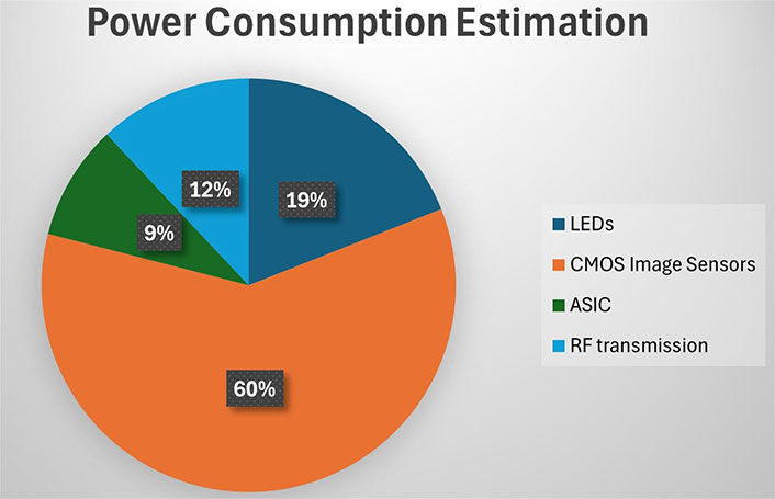
Further EC studies of integrating lithium-ion polymer and thin-film batteries promise to increase power density and reduce battery size, potentially extending operations and enhancing device capabilities [24]. Below are some important power management techniques often used in the development of ECs and other highly optimized IoT devices (Figure 6).

Power consumption estimation (source [25]). ASIC: application-specific integrated circuit; CMOS: complementary metal-oxide-semiconductor; LEDs: light-emitting diodes; RF: radio-frequency
Low-power ASIC design
ASICs are the most practical for minimizing power consumption in ECs. These ICs are custom-designed chips that perform specific functions more efficiently than general-purpose processors. In ECs, ASICs handle tasks such as image processing, data compression, and signal transmission. They are optimized for low power consumption using several important techniques:
Clock gating: clock gating is a power-saving technique that halts the clock signal to inactive sections of a chip, significantly reducing dynamic power consumption without impacting the functionality of the ASIC’s active components. It utilizes specialized clock-gating cells to manage clock distribution, targeting specific registers or units as dictated by control logic signals. This method can reduce power by up to 50%, enhancing energy efficiency in device operations [26].
Power gating: power gating extends beyond clock gating by fully disconnecting power to idle sections of the ASIC, substantially lowering static power consumption by eliminating leakage current in dormant circuits. This technique involves integrating power switches between the supply voltage and the component targeted for gating. The activation of these switches is managed by a power management unit, which sets the activity levels within the gated block to determine the appropriate times for power disconnection and reconnection. Successfully implemented, power gating can cut power leakage by more than 50% [27].
Adaptive voltage scaling: dynamic voltage scaling adjusts the supply voltage and frequency according to the workload. Lowering the voltage during less intensive operations reduces power consumption (up to 60%) while maintaining performance where needed. This method is often combined with frequency scaling to achieve optimal power-performance trade-offs. The ASIC’s power management unit dynamically adjusts the voltage and frequency based on the EC’s operating mode and performance requirements [28].
In addition to these techniques, low-power ASIC design for EC devices also involves using low-leakage transistors, such as high-k metal gate (HKMG) transistors, which minimize static power consumption. Furthermore, the ASIC’s architecture is optimized for power efficiency by implementing parallel processing, pipelining, and memory hierarchies that can reduce data movement and access energy [25].
Efficient image compression
Image compression is vital for EC power management, significantly reducing the volume of data transmitted via UHFs. High-resolution images captured by the capsule’s cameras generate large amounts of data that must be transmitted wirelessly to an external receiver. Efficient image compression algorithms are adopted to reduce this data volume, thereby conserving energy by decreasing the workload on the RF transmitter of the device.
Among the various algorithms, JPEG is the most commonly used in ECs because it effectively compresses natural scenes captured during examinations [25]. It reduces file size through discrete cosine transform, quantization, and entropy coding, effectively removing redundant information. While JPEG balances compression efficiency and image quality, PNG is an alternative for specific use cases, offering lossless compression and preserving detailed image features, which can be vital for specialized diagnostics [29].
Other advancements in image compression include exploring machine learning techniques [30] or applying region of interest (ROI) coding [31], which focuses on compressing diagnostically significant image areas. Additionally, adaptive frame rate adjustments can contribute to power savings by reducing the frequency of image captures in periods of minimal movement or activity [32]. Together, these methods enhance the energy efficiency of ECs, ensuring prolonged operation without compromising the diagnostic image quality.
ECs have significantly advanced GI diagnostics by offering a non-invasive method that integrates IoT principles. These capsules capture and transmit detailed digestive system images over the internet, enabling remote medical investigations and monitoring. In this context, it is also important to mention that current EC systems provide very short-range wireless communications due to the nature and constraints of the capsule design. Therefore, the purpose of combining IoT solutions with EC systems mainly refers to the possibility of locally integrating IoT systems and remotely sharing digitized information of the patient.
Despite their advancements, ECs face several challenges as a relatively new technology. Battery life remains a significant constraint, limiting the implementation of additional features. While some researchers like Campi et al. [33] and Zhang et al. [34] have experimented with inductive charging, current technology requires bulky external devices, sacrificing capsule mobility. Advancements in image compression, such as Google’s WebP format [35], could reduce data transmission loads, promote energy efficiency, and support EC systems to truly become self-contained IoT devices without external storage needs.
The costs associated with EC procedures also pose a barrier to widespread adoption, emphasizing the need for more cost-effective solutions. Additionally, researchers are exploring localization and tracking technologies, such as RF triangulation [36] and magnetic field sensing [37], to precisely locate the capsule, enabling more targeted diagnostics and treatment. Furthermore, improving location tracking accuracy could facilitate more detailed surgical planning and even enable cross-collaboration with robotic surgery systems. While integrating machine learning models onboard, the capsule could further optimize EC system management, adapting to individual patient situations and identifying new diagnostic patterns for various diseases.
Another limitation is that EC systems are currently unable to perform biopsies or localized treatments and lack remote navigational control, which can lead to complications such as capsule retention or travelling issues caused by GI obstructions [38].
As a solution, future advancements in EC technology could include the development of anchoring mechanisms that would allow the capsule to stop and observe specific areas of interest for extended periods. This temporal stationary ability can help with the development of a navigation and sampling system. Also, this capability could open up opportunities for targeted drug delivery and more comprehensive diagnostic imaging.
Incorporating advanced sensors into ECs, such as pH level monitors, temperature, blood detection, optical coherence tomography (OCT), electrical impedance tomography (EIT) sensors, or even biomarker detectors, could greatly enhance diagnostic precision, delivering real-time insights into the human body. These technological improvements can support the early identification and monitoring of various conditions, paving the way for nano-robotic healthcare systems capable of targeted diagnostic and therapeutic interventions.
It is also important to mention another really significant limitation of current EC system which infers the controllability and maneuverability of the system: commercial ECs are passive systems in the sense that they are swallowed into the mouth of the patient and then the trajectory of the capsule is absolutely determined by the peristaltic movements. There is no chance to slow down (or accelerate) the movement, to focus on a particular diagnostic area of interest, or to change (1) the position of the capsule and (2) the orientation of the capsule. Advances in the development of novel ECs suggested the adoption of crawling systems [39] or other technologies which are currently adopted for the movement control of catheters.
There are many other aspects that should be considered in order to improve current EC design: we mentioned before the importance of designing a robust wireless communication vs. a number of disturbances may occur in a medical environment. Enhancing the robustness of the communication protocols will inherently boost the diagnostic capability of the system (i.e. better images, higher quality of visual inspection) and the safety of the patient. Optimization of the data exchange between the EC and the visualization system or data logger will, in turn, allow exploring multiple camera configurations as well: at the moment EC usually provides only one point of view of the exploration (i.e. the frontal point of view of the capsule), however, it would be of interest to increment the visual capability with, for example, lateral cameras, especially in certain conditions were the analysis of lateral lesions is of importance.
This work presented a summary of the EC systems from the perspective of IoT technologies. By analyzing technical components, power management strategies, communication protocols, security measures, and deployment challenges, this work provided an understanding of these advanced medical tools and their role in future medical assessments.
As research progresses, EC systems have the potential to become a more powerful tool in the detection, diagnosis, and management of GI disorders. By providing real-time, comprehensive insights into the human body, these innovative IoT devices could pave the way for personalized, nano-robotic healthcare solutions capable of targeted examinations and therapeutic interventions in combination with technologies enhancing the usability and end-user interaction [40–42].
In conclusion, ECs represent a remarkable fusion of medical innovation and IoT technology, offering a patient-centric approach to GI diagnostics. While challenges remain, the ongoing advancements in EC innovations promise to revolutionize the field of gastroenterology, ultimately improving patient outcomes and quality of life.
In the future, the continued development and refinement of ECs will undoubtedly be substantial in shaping modern healthcare, leading to a new era of non-invasive, intelligent, and interconnected IoT medical devices.
AES: advanced encryption standard
ASIC: application-specific integrated circuit
CMOS: complementary metal-oxide-semiconductor
DICOM: digital imaging and communications in medicine
ECs: endoscopic capsules
ETSI: European Telecommunications Standards Institute
FCC: Federal Communications Commission
GI: gastrointestinal
HIPAA: Health Insurance Portability and Accountability Act
IoT: Internet of Things
LEDs: light-emitting diodes
RF: radio-frequency
UHF: ultra-high frequency
USD: US dollars
This work was presented in coursework form in fulfilment of the requirements for the MEng Robotics Engineering for the student Vasile Denis Manolescu under the supervision of Dr. Hamzah AlZu’bi from the School of Computer Science and the Environment, Liverpool Hope University.
VDM: Conceptualization, Investigation, Writing—original draft. HA: Supervision. ELS: Writing—review & editing.
The authors declare that they have no conflicts of interest.
Not applicable.
Not applicable.
Not applicable.
Not applicable.
Not applicable.
© The Author(s) 2024.
Copyright: © The Author(s) 2024. This is an Open Access article licensed under a Creative Commons Attribution 4.0 International License (https://creativecommons.org/licenses/by/4.0/), which permits unrestricted use, sharing, adaptation, distribution and reproduction in any medium or format, for any purpose, even commercially, as long as you give appropriate credit to the original author(s) and the source, provide a link to the Creative Commons license, and indicate if changes were made.
View: 4820
Download: 207
Times Cited: 0
Daniela Gawehns ... Matthijs van Leeuwen
Jean-Marie Grégoire ... Stéphane Carlier
