 Original Article

Affiliation:
1Department of Ultrasound, Beijing Jishuitan Hospital, Capital Medical University, Beijing 100035, China
ORCID: https://orcid.org/0000-0002-2074-4395
Affiliation:
1Department of Ultrasound, Beijing Jishuitan Hospital, Capital Medical University, Beijing 100035, China
Email: 13671053370@139.com
ORCID: https://orcid.org/0000-0002-3511-1435
Affiliation:
2Department of Pediatric Orthopedics, Beijing Jishuitan Hospital, Capital Medical University, Beijing 100035, China
ORCID: https://orcid.org/0000-0001-8666-7022
Explor Musculoskeletal Dis. 2024;2:353–359 DOI: https://doi.org/10.37349/emd.2024.00061
Received: May 16, 2024 Accepted: June 12, 2024 Published: July 31, 2024
Academic Editor: Jürgen Braun, Ruhr Universität Bochum, Germany
The article belongs to the special issue Baby Hip Sonography Worldwide: Experience, Results, and Recommendations
Aim: The aim was to review several aspects of work using ultrasound in diagnosing developmental dysplasia of the hip and analyze the application status of the Graf technique in Beijing over 15 years.
Methods: First, data on the promotion and development of the Graf technique in Beijing over the past 15 years were retrospectively analyzed. Second, data on hip ultrasound (US) screening and the effect of consecutive brace therapy were collected and analyzed. Infants were divided into subgroups according to Graf type, age at initiation of treatment, sex, and affected side.
Results: The ultrasound detection rate of developmental dysplasia of the hip (DDH) was high at Beijing Jishuitan Hospital. The total detection rate of type IIa, IIb, IIc, D, III and IV (abbreviated as: types IIa and worse) was 4.58%, and that of type IIb, IIc, D, III and IV (abbreviated as: types IIb and worse) was 1.40%. Clinicians should pay attention to DDH, and early treatment is important; therefore, it is recommended that infants and young children undergo DDH ultrasound screening as soon as possible. Our research shows that when the α angle was more than 43°, the efficacy of brace therapy was 95.95%.
Conclusions: It was confirmed that the Graf technique has greater practicality and accuracy. The Graf technique, as an important means for the early screening of DDH, has been widely recognized around the world, and it is recommended to be widely used in China to preserve future health in as many children as possible.
Developmental dysplasia of the hip (DDH) is harmful, especially in adolescence and adulthood, and may lead to gait abnormalities/claudication, differences in limb length, limited hip abduction, premature degenerative diseases of various joints, weakness of the lower extremities, scoliosis, back pain, arthritis, etc. The mildest end of the spectrum overlaps with physiologic immaturity, therefore making it difficult to determine its true incidence, which is estimated to be 1.5 to 20 per 1,000 births, depending on the demographics of the study population and the inclusion criteria [1–3]. In China, taking Beijing as an example, screening using the Graf technique to diagnose DDH has been carried out for 15 years in Beijing Jishuitan Hospital, which is the National Center for Orthopedics. During this period, He et al. [4] retrospectively analyzed the ultrasound reports of 97,332 hips in all infants who visited our hospital between January 1, 2013, and January 1, 2019, to study the efficacy of brace treatment. According to the study, the ultrasound detection rate of DDH is high: type I, 95.42%; IIa, 3.18%; IIb, 0.91%; IIc, 0.22%; D, 0.01%; III, 0.14%; IV, 0.12%. DDH was more likely to occur on the left side and in females. Once Graf IIc and worse DDH are diagnosed, bracing therapy is more effective when the child is started within 42 days of age. DDH screening is jointly performed by the Department of Ultrasound Diagnosis and Pediatric Orthopedics and mainly targets infants born in and around Beijing. This work led to the widespread roll-out of DDH screening across the city and country. Reinhard Graf was invited several times to give lectures on his technique, which is the internationally recognized diagnostic method for DDH.
The main purpose of this article is to summarize several aspects of work using ultrasound in diagnosing developmental dysplasia of the hip and analyze the application status of the Graf technique in Beijing over 15 years.
Since 2009, we have performed DDH ultrasound on 70,000 children, and we have always adhered to the philosophy regarding the recommendation of clinical examination; however, clinical examination is subjective and cannot solve this problem. In 2020, our center undertook the clinical innovation project of the Beijing Hospital Administration (research on the standardization of ultrasound diagnosis of DDH in infants) to promote the Graf technique to more than 20 units at the grassroots level and provide a “quick referral” for children. According to statistics, during a one-year time period, more than 10,000 infants who underwent hip screening in primary hospitals had more than 500 hips suspected of DDH and more than 300 children were referred.
To obtain the distribution characteristics, we divided the infants into subgroups according to the different Graf types (I–IV), sex, affected side, and age. It was ensured that clear Graf standard sectional images were collected while the examined infants were completely relaxed.
For hips classified as Graf type IIc or D, in accordance with the standard of Atalar et al. [5] success was defined as follows: ultrasonography showing that the femoral head and acetabulum were well aligned, an α greater than 60°, the presence of an X-ray pelvic flat piece reflecting good alignment of the femoral head and acetabulum, or an AI less than 25°. For hips classified as Graf type III or IV, success was defined as follows: medial hip US showing that the dislocated femoral head had achieved concentric reduction. According to the Graf method, the US results indicate a change in the Graf type from IIc, D, III, and IV to type IIb or I, and the X-ray pelvic flat piece reflects good alignment of the femoral head and acetabulum.
In the course of treatment, if ultrasonography or radiography from three consecutive reviews indicate that the femoral head cannot be reduced, the acetabulum development is not further improved, or there are complications such as avascular necrosis of the femoral head and femoral nerve paralysis, then the treatment of the sling or brace has failed and is considered ineffective.
SPSS 26.0 was used for the statistical analysis. The statistics analysis was mostly descriptive with most data presented as absolute numbers and rates (percentages). In the analysis of the efficacy of brace therapy, the chi-square test or exact probability test was used for comparisons between count data groups. We divided the infants into subgroups according to the different Graf types (I–IV), sex, affected side, and age to compare the efficacy between different groups and whether the difference was statistically significant. Bilateral tests were used for all tests, and P < 0.05 was considered to indicate statistical significance.
According to a large amount of data, we confirmed that the Graf technique is widely used in training, teaching, quality control, and application in Beijing and is gradually advancing to standardization.
Previous studies of all infants examined from January 2013 to January 2019 have concluded that among the 48,666 infants examined, the detection rates were as follows [4]: Graf type I, 95.42%; IIa, 3.18%; IIb, 0.91%; IIc, 0.22%; D, 0.01%; III, 0.14%; and IV, 0.12%. So, the detection rate of Graf type IIa and worse was 4.58%, and that of Graf type IIb and worse was 1.40%.
To evaluate the efficacy of brace therapy in infants with Graf IIc and worse, we followed up patients from 2013 to 2018 and found that the overall early treatment response rate to DDH brace treatment was 74.70%, and the early treatment effective rate curve over time is shown in Figure 1.
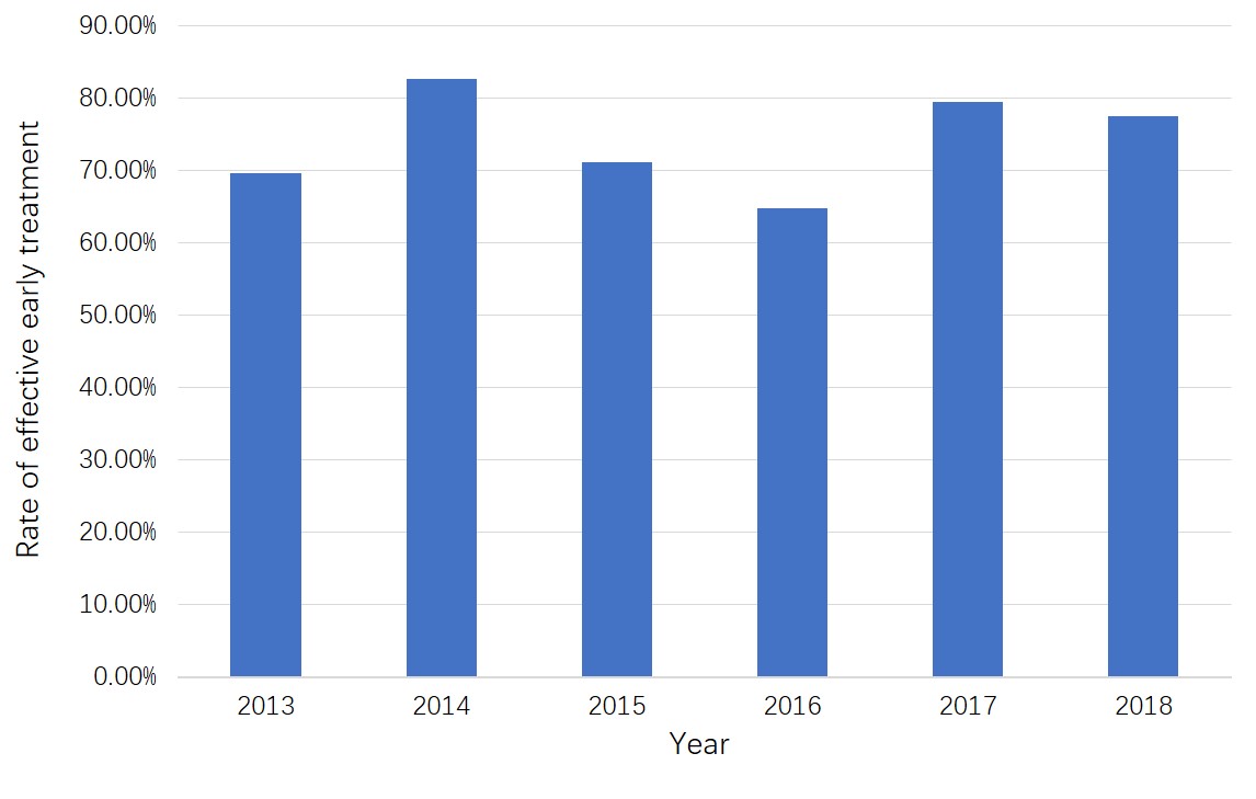
Rate of effective early treatment in infants with Graf type IIc and worse from 2013 to 2018
We further followed up with a group of patients with hip joints classified as Graf IIa and Graf IIb from January 1, 2021, to January 1, 2022. A total of 754 infants were retrospectively studied, and all the patients with alpha angles less than 60 and more than 50 degrees were included. The age with standard deviation for the first visit was 79.5 ± 40.3 days, the minimum age was 6 days, and the maximum age was 209 days. A total of 1,291 hips were included, 126 (16.70%) left hips were affected, 91 right hips (12.10%) were affected, 537 bilateral hips (71.20%) were affected, 607 infants (80.50%) were female, and 147 infants (19.50%) were male (Table 1). We wanted to study these children because Graf ultrasound technology allows them to be followed throughout the recovery process, and in most cases, the hip becomes stable and mature with a brace or just a follow-up. Ultrasonography was performed, and at the same time, the pediatric orthopedic outpatient review was carried out, to observe the effect of brace treatment, the highest number of follow-up visits was 10 for the same child. Results of the analysis of outcomes in this group of patients are planned to be published later.
Characteristics of infants with Graf IIa and IIb (the patients with an alpha angle less than 60 and more than 50 degrees) according to different affected sides and gender during January 1, 2021 to January 1, 2022
| Classes | Infants | Detection rates (%) |
|---|---|---|
| Affected side | ||
| Left | 126 | 16.70% |
| Right | 91 | 12.10% |
| Bilateral | 537 | 71.20% |
| Gender | ||
| Female | 607 | 80.50% |
| Male | 147 | 19.50% |
| Total | 754 | 100.00% |
To study the efficacy of brace therapy, we analyzed the results of the treatment in children with complete follow-up records. Brace therapy was applied to 423 affected hips (Graf type IIc, 202 hips; Graf type D, 17 hips; Graf type III, 118 hips; and Graf type IV, 86 hips). For follow-up efficiency, US, X-ray, and clinical examination were used. Based on the success of early treatment with the brace, the patients were divided into “effective” and “ineffective” groups. The statistical results showed that as the α angle increased (P < 0.05), the β angle decreased (P < 0.05). When the α angle was more than 43°, the efficacy of early treatment was 95.95%.
In 2019, according to the International Interdisciplinary Consensus Committee on DDH Evaluation (ICODE), neonatal clinical examination may detect only hip instability but not acetabular dysplasia; ultrasound, on the other hand, is more sensitive and specific for diagnosing DDH [6]. In 2023, the British Society for Children’s Orthopaedic Surgery (BSCOS) recommended universal hip ultrasound for screening for DDH in infants up to 3 months of age [7]. The ICODE makes it clear that the Graf technique is the preferred method for ultrasound in the diagnosis of early DDH and needs to be performed by a trained and certified sonographer [6]. The Graf technique can be used to correlate the pathology and severity of acetabular dysplasia with the age of the child to provide a specific diagnosis and treatment to prevent overtreatment. The BSCOS consensus also states that the Graf technique should be used as the standard ultrasound screening method for DDH.
Ultrasound can be used to observe hip structure noninvasively, dynamically, and comprehensively and is helpful for providing a basis for the clinical diagnosis of DDH, classification evaluation, treatment plan formulation, and prognosis evaluation. The Graf technique has been widely recognized in many parts of China, including in Beijing, Tianjin, Shanghai, and Yangzhou, and abroad as an important means for early screening of DDH. It is recommended that this technique be widely used in China to save more children.
Our center holds at least two DDH ultrasound training courses every year, training a total of 2,000 students and enrolling 200 ultrasound doctors from hospitals all over China. These trainees learn through observation and hands-on training during a three-month period. After the outbreak of COVID-19, enthusiasm for learning the Graf technique did not decrease across the country, and online courses could help more students learn the Graf technique. In October 2021, 70,000 students participated in the Jishuitan Forum and DDH ultrasonic examination method course. In 2023, the 23rd National Conference on Ultrasonic Medicine of the Chinese Medical Association invited Graf to give a lecture demonstrating the Graf technique to 2,500 participants in Beijing. As the technical backbone, these doctors can improve the DDH diagnosis and treatment level through training and vigorously carry out DDH screening after returning to their units to benefit more children.
He et al. [8] previous research revealed that the earlier the treatment is, the better the treatment efficacy, especially for Graf type IIc; conservative treatment of children with Graf type IIc and worse is best started within 42 days of life, and the treatment response is best at this time. Among the infants screened in our center, the detection rate of Graf type IIc and worse was as high as 4.9% [4]. If these children cannot be screened early or receive standardized diagnosis and treatment in a timely manner, the optimal treatment opportunity will be missed, which will cause serious consequences and complications in the late stage. Therefore, we recommend that DDH hip screening be carried out when conditions permit and emphasize the role of ultrasound in follow-up evaluation during treatment.
The diagnosis and treatment of patients in the pediatric orthopedic department are at the highest level in China, with a large number of patients treated and good therapeutic effects. Lyu et al. [9], in a study of the efficiency of different types of braces, reported that the Tübingen splint should be the preferred treatment option for children older than three months and for those with severe forms of DDH, such as Graf type III and IV DDH, who are younger than six months at the time of diagnosis. The Tübingen hip flexion splint is a valid alternative to the Pavlik harness for older infants and those with more severe DDH.
In recent years, to improve the efficiency of ultrasound diagnosis and help more children, we developed the artificial intelligence three-dimensional ultrasonic imaging automatic diagnosis system to make more accurate diagnoses. The Graf technique is currently the most commonly used method for diagnosing DDH in infants; this technique has been proven to be reliable and has a good correlation with other methods [10]. Chen et al. [11] developed a deep learning-based computer-aided framework for DDH diagnosis to minimize manual intervention in the diagnosis process. The framework can perform fully automated standard plane detection and angle measurements for Graf type I and type II hips. In early studies, the following methods were adopted: 1,051 US images and 289 US videos of Graf type I and type II hips were used to evaluate the performance of the proposed framework. In static mode, the mean absolute errors of the α and β angles are 1.71° and 2.40°, respectively, and the classification accuracy is 94.71%. In dynamic mode, the mean absolute errors of the α and β angles are 1.97° and 2.53°, respectively, the classification accuracy is 89.51%, and the running speed is 31 fps. The experimental results demonstrate that our fully automated framework can accurately perform standard plane detection and angle measurement of an infant’s hip at a fast speed, showing great potential for clinical application. He et al. [12]. concluded that it is feasible to use multiplanar measurements of infant hips with three-dimensional ultrasonography.
Regardless of the type of orthosis, the mechanism of action is the same; all mechanisms can provide and sustain a stable, concentrically reduced hip joint at the earliest possible age [13, 14]. Our previous studies on the efficacy of braces in children in Beijing confirmed that if undiagnosed and untreated subluxation and dislocation can be treated conservatively to obtain good results, degenerative joint disease can be avoided.
In the next step, our team will continue to follow these children and record whether they received brace therapy to analyze the effect of the brace on their development of stable and mature hip joints. It would be more important to perform a prospective study where we try to determine the characteristics of children with an alpha angle ≥ 50° who need to wear braces to prevent overtreatment.
In conclusion, since Graf introduced ultrasonographic classification of the hip joint in 1980, the role of ultrasonography in diagnosis and screening has increased. It was confirmed that the Graf technique has greater practicality and accuracy. The Graf technique, as an important means for the early screening of DDH, has been widely recognized around the world. As the study reports, patients whose α angle was more than 43° are recommended to receive brace therapy as soon as possible for the best effects. It is recommended to be widely used in China to preserve the future health of as many children as possible.
The innovation and significance of this study mainly includes the following points:
Provides a review of the Graf technique’s application in Beijing over 15 Years, offering insights into long-term trends and effectiveness.
Promoting the Graf technique to grassroots-level units and providing “quick referral” is an innovative approach to improving access to care.
Highlights the importance of training healthcare professionals in the Graf technique, which has been done through regular courses.
Suggests the need for further prospective research to determine the characteristics of children who need brace therapy, preventing overtreatment.
The development of an artificial intelligence three-dimensional ultrasonic imaging automatic diagnosis system is an innovation that can enhance diagnostic accuracy and efficiency.
In summary, this study is significant as it not only reviews the application of the Graf technique but also contributes to the standardization, training, and technological advancement in the detection and early treatment of DDH.
DDH: developmental dysplasia of the hip
US: ultrasound
JH: Conceptualization, Data curation, Investigation, Writing—original draft. TC: Validation, Writing—review & editing, Supervision. XL: Conceptualization, Writing—review & editing. All authors read and approved the submitted version.
The authors declare that they have no conflicts of interest.
This study was approved by the institutional review board (IRB) of Beijing Jishuitan Hospital (code: 201805-09), (code: 202004-87), and (code: 202104-29) complies with the “Declaration of Helsinki”, “International Ethical Guidelines for Human Biomedical Research”, and “Designers’ Biomedical Research Ethical Review Measures”.
The requirement of informed consent has been waived by IRB as this study was a retrospective study.
Not applicable.
Corresponding authors can be contacted for relevant study data.
We thank the Beijing Jishuitan Hospital Research Fund-Medical Industry Enterprise Cross-cultivation Project [YGQ-202303]; Beijing Hospitals Authority Clinical Medicine Development of special funding [YGLX202319] support, for providing special funding support in the study design; in the collection, analysis, and interpretation of the data; in the writing of the report; and in the decision to submit the paper for publication.
© The Author(s) 2024.
Copyright: © The Author(s) 2024. This is an Open Access article licensed under a Creative Commons Attribution 4.0 International License (https://creativecommons.org/licenses/by/4.0/), which permits unrestricted use, sharing, adaptation, distribution and reproduction in any medium or format, for any purpose, even commercially, as long as you give appropriate credit to the original author(s) and the source, provide a link to the Creative Commons license, and indicate if changes were made.

View: 2663
Download: 19
Times Cited: 0
Hakan Ömeroğlu ... Feza Korkusuz
Maurizio De Pellegrin ... Nicola Guindani
Beat Dubs
Beat Dubs
Giovanna Galvão Braga Motta ... Alexandre Francisco de Lourenço
Nicholas Birkett ... Claudia Maizen
Tanja Kraus, Catharina Chiari
Konstantinos Chlapoutakis ... Maria Raissaki
