
Abstract
The integration of digital technologies like CAD/CAM systems and intraoral scanners is transforming the field of dentistry, providing numerous benefits for both patients and dental professionals. This case study explores the significance and benefits of dental CAD/CAM technology in revolutionizing the dental industry, focusing on the creation of personalized dental restorations with precision and speed. The study emphasizes the applications of CAD/CAM technology and showcases the state-of-the-art Medit i700 intraoral scanner for detailed dental impression scanning. By adopting these advanced technologies, dental practices can save time, enhance patient satisfaction, and reduce costs in the long run. The methodology involved systematic digital assessments, planning, and execution to address cosmetic concerns, resulting in a successful transformation of a patient’s smile using e.max® CAD/CAM technology and Medit i700 scanner. The case study highlights the importance of patient preference, efficiency, and accuracy in digital dentistry, demonstrating the potential for providing personalized and effective solutions for aesthetic dental concerns. Embracing digital advancements and patient-centered care is crucial for delivering high-quality and innovative solutions in modern aesthetic dentistry.
Keywords
CAD/CAM, smile design, customized ceramic models, digital prosthesis manufacturingIntroduction
Today’s dental practice makes use of a variety of cutting-edge tools, such as scanners, printers, and milling machines, in order to perform aesthetic procedures that enhance the overall look of a patient’s face and smile. Whether it involves making small adjustments to improve minor aesthetic flaws or completely transforming more significant cosmetic aspects, all of these elements play a vital role in creating a beautiful smile. While in the past, virtual smile design was achieved through the use of image editing software and traditional or virtual wax models, the current process of creating customized patient models demands a higher level of proficiency in computer technology [1–4]. As digital dentistry continues to advance, it is significantly changing the way dental treatments are administered and ultimately resulting in better outcomes for patients. Dental CAD/CAM systems are essential in this transformation, allowing for more accurate dental restorations and enhancing the overall patient experience [5–12]. With the advancement of technology, dental professionals can now use CAD/CAM systems for various applications, from creating custom crowns and bridges to designing orthodontic appliances. These systems not only simplify the fabrication process but also produce more precise and aesthetically pleasing results. Looking forward, the future of dental CAD/CAM systems shows promise with advancements in artificial intelligence and 3D printing technology offering new opportunities for personalized and efficient dental care. These innovations will enable dental professionals to provide better care and cater to the unique needs of each patient. In conclusion, the integration of digital technologies like CAD/CAM systems is transforming the field of dentistry, providing benefits for both patients and dental professionals [13–17]. By embracing these technologies and staying informed about the latest developments, the future of dental care looks brighter than ever. Besides, digital dentistry is a vital component of modern dental practice, incorporating advanced technology like intraoral and facial scanners, 3D printers, and milling. Integration of these systems is crucial for achieving patient satisfaction and enhancing facial aesthetics. Virtual smile design has evolved from 2D simulations to 3D digital models, revolutionizing the creation of personalized patient models. Lithium disilicate ceramics have become a popular choice for aesthetics and strength in dental restorations. Digital impressions through intraoral scanners save time, improve accuracy, and enhance communication with patients and labs. While digital techniques have numerous advantages, they require expertise and investment in equipment. In summary, digital dentistry is transforming the field with its precision and efficiency in creating beautiful smiles. This case study delves into the significance and benefits of dental CAD/CAM technology in revolutionizing the dental industry [16–19]. The exploration begins with an in-depth look at the importance of digital transformation in dentistry and highlights how this technology allows for the creation of personalized dental restorations with utmost precision and speed. Additionally, the study showcases the diverse applications of CAD/CAM technology across various sectors and introduces the state-of-the-art Medit i700 intraoral scanner, which offers detailed dental impression scanning. The scanner’s notable features include high-quality scans, rapid scanning speeds, powder-free scanning, immediate feedback, and versatility in use.
The study emphasizes the numerous advantages of integrating these advanced technologies into dental practices, such as time-saving benefits, enhanced patient satisfaction, and long-term cost reduction. It ultimately asserts that the adoption of CAD/CAM technology and intraoral scanners like the Medit i700 is essential in modern dentistry to provide precise, efficient, and cost-effective dental care. The case study aims to explore the feasibility of incorporating digitally created veneers through computer-based methods, showcasing the potential for enhanced outcomes in dental treatments.
Case report
The case study conducted at the Faculty of Dentistry at Damascus University from August 1, 2020, to January 2, 2022, focused on utilizing digital technology to improve the cosmetic appearance of a 23-year-old female patient’s smile. The patient expressed dissatisfaction with the gap between her upper incisors and sought a non-invasive solution. Upon examination, orthodontic treatment was initially recommended but the patient opted for a more immediate and aesthetic solution. It was proposed to create four porcelain veneers using e.max® CAD/CAM technology to close the gap and enhance the overall appearance of her smile. The Medit i700 scanner was used to capture a detailed 3D model of the patient’s upper jaw through an orofacial scan. This innovative approach allowed for the precise design of the veneers, ensuring a custom fit and natural look. The methodology employed in this case study involved a systematic approach to utilizing digital technology to address the cosmetic concerns of the patient. Various stages were followed to ensure a comprehensive assessment, treatment planning, and execution of the treatment. Initially, extraoral and intraoral photographs were taken to assess the patient’s condition and plan the treatment. Subsequently, intraoral scans of the upper and lower jaws were performed using the Medit i700 scanner to capture detailed 3D models. Digital smile design was carried out to visualize the desired outcome and plan the treatment accordingly. Tooth preparation was then performed, followed by another intraoral scan to ensure accuracy and precision. The color of the veneers was digitally selected using the software’s color selection feature to achieve an accurate color match. The porcelain veneers were then cemented to the teeth using a bonding protocol, ensuring proper alignment and fit. Throughout the treatment process, attention was paid to detail and accuracy, with regular checks and tests conducted to ensure the success of the treatment. Pre- and post-treatment photographs were taken to document the results and showcase the transformation achieved through digital technology. Overall, the method employed in this case study emphasizes the importance of thorough assessment, precise planning, and detailed execution when utilizing digital technology in cosmetic dentistry. By following a systematic approach and incorporating innovative digital tools, personalized and effective solutions can be provided to address patients’ aesthetic concerns. The digital transformation of the treatment plan not only met the patient’s cosmetic goals but also highlighted the advancements in digital dentistry. The study demonstrated the potential of digital technology in creating personalized and effective solutions for aesthetic dental concerns.
The following stages were involved in the study:
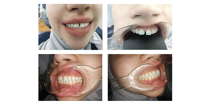
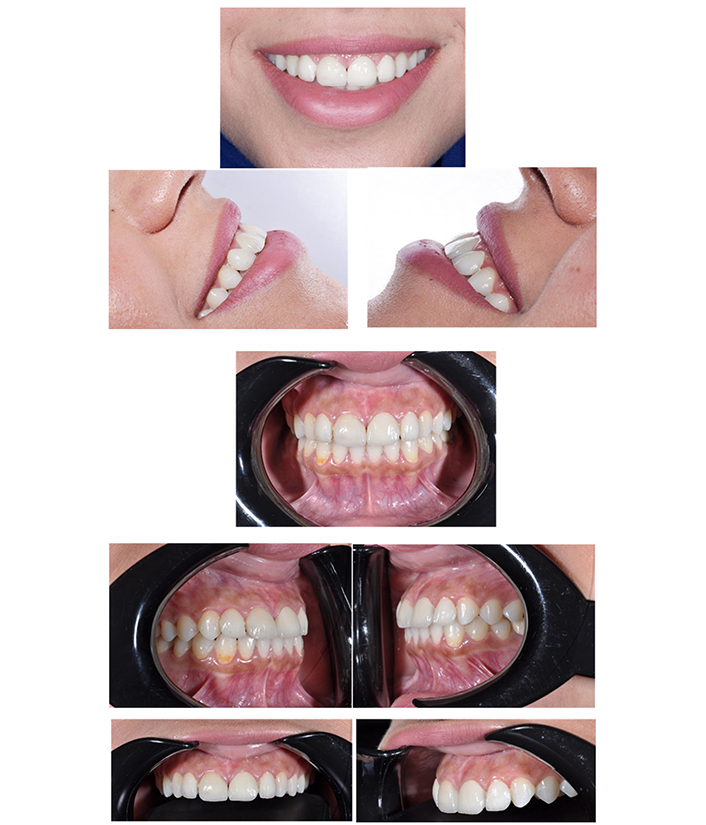
Various extraoral and intraoral photographs were taken in different positions (Figure 1), including frontal views with and without a smile, lateral view of the facial profile, and intraoral views with retractors. These images helped in assessing the patient’s condition.
Before starting the treatment, an intraoral scan of both the upper and lower jaw was performed in a lateral occlusal position (Figure 2) using the Medit i700 device.
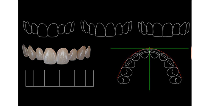
Digital smile design (Figure 3) was carried out to plan the desired outcome.
Tooth preparation began by using an appropriate bur sequence, with the finishing line placed above the gingival level.
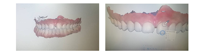
After the tooth preparation was completed, an intraoral scan was performed again. The color of the veneers was chosen digitally using a color selection feature in the software (Figure 4), ensuring accurate color matching for each tooth area.
The porcelain veneers were then cemented to the teeth after thorough testing and ensuring no issues. Adjacent teeth were isolated, and the bonding process followed the usual protocol for porcelain veneers.
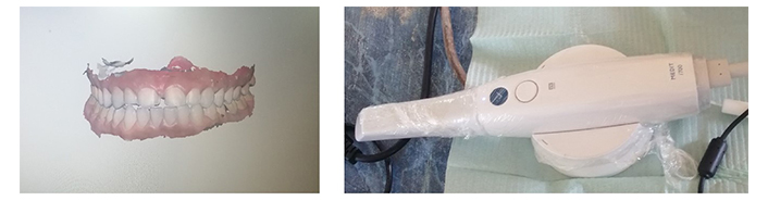
The scanner of the upper and lower jaws with the occlusion using Medit i700 scanner
Treating the veneer involves using 10% hydrofluoric acid to etch the inner surface for 20 seconds, followed by washing with water. Silane is then applied for a minute, followed by a layer of bond and cement, specifically a light-cured resin cement. Treating the abutments involves applying 37% phosphorous acid to the entire labial surface of the teeth for 30 seconds, followed by washing with water. A bonding material is then applied before carefully placing the veneers on the teeth, ensuring proper alignment. After light curing for two seconds, any excess material is removed before allowing sufficient time for curing. Each surface is cured for 40 seconds. Finally, it is recommended to take pre- and post-treatment photographs, both intraoral and extraoral, to document the results (Figure 5).
Discussion
The utilization of digital technology in the field of dentistry has revolutionized the way aesthetic concerns are addressed, as demonstrated in this case study. By incorporating e.max® CAD/CAM technology and the Medit i700 scanner, a personalized treatment plan was developed to improve the cosmetic appearance of the patient’s smile. The use of digital smile design allowed for precise and detailed visualization of the expected outcome, ensuring patient satisfaction and enhanced communication between the dentist and the patient. The case study highlighted the importance of considering patient preferences and desires when planning treatment options. Despite the initial recommendation of orthodontic treatment, the patient’s preference for a more immediate and aesthetic solution was respected. This demonstrates the importance of offering a range of treatment options and involving the patient in the decision-making process. The successful outcome of this case study emphasizes the potential of digital technology in improving the efficiency and accuracy of dental treatments. The ability to capture detailed 3D models of the patient’s teeth and design customized veneers using CAD/CAM technology ensures a high level of precision and fit. Additionally, the digital color selection feature allowed for accurate color matching, enhancing the natural look of the veneers. Overall, this case study showcases the transformative impact of digital technology in dentistry, particularly in addressing cosmetic concerns and enhancing patient satisfaction. As digital dentistry continues to evolve, it is likely to play an increasingly significant role in providing personalized and effective solutions for patients seeking aesthetic improvements.
Conclusions
Utilizing CAM/CAD technology in dentistry revolutionizes the design and production of dental prosthetics and devices, resulting in precise and efficient outcomes. This innovative approach not only reduces errors but also enhances patient comfort, fit, and overall aesthetics. The case study showcased the successful application of digital technology in addressing a patient’s desire for an improved smile. Through meticulous assessment, planning, and execution, the treatment led to a remarkable transformation in the patient’s appearance. By utilizing cutting-edge tools like the Medit i700 scanner and e.max® CAD/CAM technology, personalized porcelain veneers were created, achieving a natural and aesthetically pleasing result. Patient involvement and preference were vital aspects highlighted in the study, as the patient opted for a non-invasive solution over orthodontic treatment. Emphasizing patient-centered care and offering a range of treatment options proved instrumental in achieving favorable outcomes. Moreover, the study underscored the efficiency and accuracy that digital technology brings to dental procedures. Capturing detailed 3D models, designing custom veneers, and digitally selecting colors for precise matching all contributed to the success of the treatment. This emphasizes the transformative potential of digital dentistry in providing tailored solutions for patients seeking aesthetic enhancements.
Ultimately, the systematic approach and integration of digital technology demonstrated in this case study exemplify a forward-thinking strategy for future dental procedures. Embracing digital advancements and prioritizing patient preferences can further enhance the delivery of high-quality and innovative solutions in aesthetic dentistry.
Declarations
Author contributions
ZD and AD: Conceptualization, Data curation, Formal analysis, Funding acquisition, Resources, Writing—original draft, Writing—review & editing. MA: Conceptualization, Data curation, Formal analysis, Writing—original draft, Writing —review & editing. CA: Data curation, Writing—review & editing.
Conflicts of interest
The authors declare that they have no conflicts of interest.
Ethical approval
This study is approved by the ethics committee at Damascus University.
Consent to participate
Informed consent to participate was obtained from the relevant participant.
Consent to publication
Informed consent to publication was obtained from the relevant participant.
Availability of data and materials
The data and materials that support the findings of this study are available from the corresponding author upon reasonable request.
Funding
The study is funded by Damascus University (https://www.damascusuniversity.edu.sy/index.php?lang=2), and International University for Science and Technology (https://iust.edu.sy/en/). The funders had no role in study design, data collection and analysis, decision to publish, or preparation of the manuscript.
Copyright
© The Author(s) 2024