Original Article

Affiliation:
Department of Restorative Dentistry, Kornberg School of Dentistry, Temple University, Philadelphia, PA 19140, USA
Email: salwa.mekled@temple.edu
ORCID: https://orcid.org/0009-0001-1638-9159
Affiliation:
Department of Restorative Dentistry, Kornberg School of Dentistry, Temple University, Philadelphia, PA 19140, USA
Affiliation:
Department of Restorative Dentistry, Kornberg School of Dentistry, Temple University, Philadelphia, PA 19140, USA
Affiliation:
Department of Restorative Dentistry, Kornberg School of Dentistry, Temple University, Philadelphia, PA 19140, USA
Affiliation:
Department of Restorative Dentistry, Kornberg School of Dentistry, Temple University, Philadelphia, PA 19140, USA
Affiliation:
Department of Restorative Dentistry, Kornberg School of Dentistry, Temple University, Philadelphia, PA 19140, USA
Affiliation:
Department of Restorative Dentistry, Kornberg School of Dentistry, Temple University, Philadelphia, PA 19140, USA
Explor Med. 2024;5:566–573 DOI: https://doi.org/10.37349/emed.2024.00240
Received: April 25, 2024 Accepted: June 14, 2024 Published: August 01, 2024
Academic Editor: Gaetano Isola, University of Catania, Italy
The article belongs to the special issue Biomaterials and Biomarkers in Dentistry: Up to Date
Aim: To evaluate the fracture resistance of printed crowns with two different designs.
Methods: Forty restorations (n = 20/group) were fabricated using resin matrix ceramic (VarseoSmileTM) with different designs as follows: group 1: overlay restoration with chamfer margin; group 2: overlay restoration with chamfer margin located 3 mm above the gingiva. Restorations were bonded to resin dies using 3M RelyX Luting Cement. Compression forces were applied with a 4 mm diameter steel bar at the midline fissure of each crown using the universal testing machine. Restorations were loaded until fracture, and fracture resistance at maximum load was recorded. Statistical analysis: A t-test was used for statistical comparison between groups.
Results: No difference in the fracture resistance was detected for both groups.
Conclusions: Different designs of composite resin crowns fabricated using CAD/CAM techniques can be used, however further research is needed to assess the clinical outcomes.
The use of computer-aided design/computer-aided manufacturing (CAD/CAM) technology has extensively expanded in restorative dentistry [1, 2]. The introduction of CAD/CAM was challenging and has seen several developments since the 1980s to reduce the high cost, decrease operation time, and ease system manipulation. CAD/CAM has played an important role in advancing fixed indirect restorations, and removable appliances to restore patients’ oral function [3]. Nowadays clinicians can perform varieties of procedures with full digital workflows [4–6]. The development of digital dentistry reduces human error, and it aims to fabricate restorations faster, more accurately, and with predictable outcomes [7, 8]. Moreover, clinicians can manufacture a large variety of chairside CAD/CAM restorations with different types of ceramic without the assistance of the dental technician [9].
Conservative tooth preparations limit the removal of the defective tooth structure and aim to maintain the remaining intact tooth structure, while ensuring that the preparation has the appropriate space for the material needed [10]. Traditional full coverage crowns are not a conservative approach because studies have shown that the traditional tooth preparation needs to remove approximately 24–70% of the existing tooth structure, and therefore it may compromise the longevity of the tooth [11, 12]. Conservative tooth preparations reduce the trauma to the pulpal tissue and avoid subgingival finish lines that may irritate periodontal tissues [13, 14].
Furthermore, partial coverage crowns have also been used as an alternative to restore occlusal wear avoiding traditional full coverage crowns and have been shown to successfully restore masticatory function [15, 16]. An overlay restoration is a type of partial restoration that only covers the occlusal surface of the tooth [17]. An overlay restoration can also be found with other names in the literature such as occlusal veneers, tabletops, or simply as partial restoration [18–20]. These restorations preserve tooth structure however, the used material should be strong enough to resist masticatory forces [19]. Resin cement and dental adhesives can strengthen the remaining tooth structure and therefore a conservative restoration such as an overlay preserving as much of the intact tooth structure is ideally indicated [21–23]. Recently, ceramic-infiltrated hybrid composites have higher mechanical characteristics, thus have been used to 3D print definitive restorations such as single crowns, inlays, onlays, tabletops, and veneers [24, 25].
VaresoSmileTM is a resin matrix ceramic (RMCs) that contains both methacrylic ester matrix and ceramic fillers [24]. Limited amount of research has compared different crown designs using VarseoSmileTM Crown. Our study investigates the fracture resistance of printed crown restorations with two different designs. The null hypothesis of the present study is that there is no difference in fracture resistance between ceramic-infiltrated hybrid composite crowns fabricated with two different designs.
Two typodonts (1560 Dentoform, Columbia Dentoform, Lancaster, PA, USA) mandibular right first molar teeth were prepared for full coverage crown (group 1), and overlay restorations (group 2), respectively for each type of preparation with 1.0 mm occlusal reduction with chamfer, and chamfer 3 mm above the gingival level, and circumferential reduction by 1.0 mm. The preparations had an axial wall taper of 6 to 8 degrees. The amount of reduction compensates for the minimal thickness of the selected crown material according to the manufacturer’s guideline [24, 25].
All the preparations were done using a flat-end long tapered diamond (Bur # TF-14, ISO 172/023, Mani Inc., Japan) with a highspeed contra-angle handpiece Ti-Max Z900L (NSK, Nakanishi Inc., Tochigi, Japan). A putty matrix was used to ensure the standard measurements of all the preparations. An optical scanner (E1 scanner, 3Shape, København, Denmark), was used to scan both resin typodonts. The scanned models were fabricated with a 3D printer (Asiga MAXTM; Asiga, Sydney, Australia) using model resin materials (Asiga DentaMODEL, Asiga, Sydney, Australia) [26].
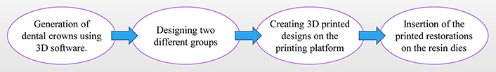
A virtual design was created for the two different designs. Forty restorations (n = 20/group) were fabricated using Exocad software (Exocad GmbH, Darmstadt, Germany) in 3D, according to ISO 4949-2019 (Figure 1). The data was then transferred to standard tessellation language (STL) files and transmitted to a 3D printer software program to determine printing procedures. Resin dies were fabricated to match the preparations using a model resin (Formlabs, Somerville, MA, USA).
The restorations were polished with pumice stone and polishing compound according to the manufacturer’s guidelines to achieve better surface quality. All restorations were sandblasted with 50 µm aluminum oxide for 10 seconds. Restorations were bonded to resin dies using RelyX Automix Luting Plus Cement (3M, ESPE). Compression load of 50 N was placed at the midline fissure of each sample with a 4 mm diameter steel bar using the universal testing machine (Instron 5569 Universal). Restorations were loaded until fracture, then fracture resistance at maximum load was recorded.
The data were analyzed using two sample t-test. All statistical analyses were performed using a statistical software program (IBM SPSS Statistics, v25, Armonk, NY, USA). Analyses were used to compare the average compression load and compression extension between full coverage crown design (group 1) and overlay design (group 2).
Table 1 displays the results of the present study. The mean compression load, and the mean compression extension of traditional crown and overlay restorations with 3 mm above the gingiva for the mandibular first molar did not significantly differ.
Compressive load and compressive extension between crown group and overlay group that indicates no significant differences (p > 0.05)
| Group | Mean | SD | P-value(Two-sample t-test) | Median | P-value(Mann-Whitney U test) |
|---|---|---|---|---|---|
| Compressive load | |||||
| Crown group | 1,648.2 | 333.4 | 0.1090 | 1,800 | 0.1285 |
| Overlay group | 1,457.9 | 499.0 | 1,500 | ||
| Compressive extension | |||||
| Crown group | 2.5 | 0.4 | 0.1178 | 2.5 | 0.0561 |
| Overlay group | 2.0 | 1.3 | 2.2 | ||
Our study found that the mean compressive load for full coverage crown (group 1) was 1,648.2 N (± 333.4), while the mean compressive load for overlay restorations (group 2) was 1,457.9 N (± 499). The mean fracture extension for full coverage crown (group 1) was 2.5 mm (± 0.4), while the mean fracture extension for overlay was 2.0 mm (± 1.3).
A Mann-Whitney U test was performed to compare compressive loads and fracture extension between the full coverage crown group (group 1) and the overlay restorations (group 2). The present study used software for statistical analysis (IBM SPSS Statistics, v25, Armonk, NY, USA), focusing on mean compressive load and compressive extension between different groups.
Detailed information comparing different groups is in Table 1. In both analyses, the p-value was > 0.05. Therefore, there was not enough evidence to reject the null hypothesis.
Partial coverage restorations have become a popular treatment option in recent years and clinical studies evaluating them are demonstrating promising results. A controlled clinical trial evaluated ceramic partial coverage restorations for posterior teeth on 22 patients for 5.5 years concluding with an 88.8% survival rate [27]. A recent systematic review evaluating the longevity of partial ceramic restorations found survival rates of 91–100% at 2–5 years and 71–98.5% for more than 5 years, and concluded that ceramic partial restorations appear to be a reliable option for posterior teeth regardless of the follow-up duration [28]. Zirconia partial restorations covering the entire surface have been widely described in case reports with different designs such as following the occlusal anatomy, flat occlusal surface, with finish margin located at different heights, or even without finish margin [29, 30].
Full crown preparations can cause the loss of a large amount of the remaining tooth structure after pathology or endodontic treatment [31]. Recent research investigates partial restorations that preserve more tooth structure than traditional full crowns [13, 32]. The fracture load may be affected by factors such as material type, the restorative design, and the choice of cementation material [33, 34]. Therefore, the occlusal reduction in this present study was 1 mm according to the minimal thickness needed for VaresoSmileTM crowns manufacturer manual. RelyX luting cement was used to provide a high fracture resistance and great wear resistance [35].
The use of CAD/CAM resin composite blocks has been increasing recently [36]. CAD/CAM crowns require sufficient axial wall thickness, and sufficient crown height. There is limited research that evaluates the mechanical properties of 3D-printed resin used for full coverage crowns [34]. In vivo studies show a low success rate in the premolar area. A three-year retrospective cohort study of 547 CAD/CAM crowns showed complications, such as retention loss and fracture [34]. The glass-ceramic CAD/CAM crowns were more prone to irreparable fracture [37]. Contributing factors that lead to loss of retention of the crown include bonding technique, contamination with saliva [38], and crown fit [39]. Adding Zirconia and micro fillers to 3D-printed composite resin would increase strength and surface roughness compared to unmodified composite resin [33]. Adding micro fillers or pre-polymerized resin will increase the filler content and improve mechanical properties [40]. In our study, we used “RMCs” that contain both methacrylic ester matrix and ceramic fillers.
The present study found no significant difference in the compression load required to fracture the crowns printed with the two different designs, thus underscoring the importance of a comprehensive understanding of the mechanical behavior of different crown overlay designs. Our study found that the compression load needed to fracture composite resin crown and overlay restorations were 1,800 N and 1,500 N respectively. Although there was no significant difference between full coverage crown and overlay restoration, our results were higher than the maximum biting forces in the posterior area (500–900 N). Our results agreed with several previous studies that revealed similar results as forces between 1,495 and 2,303 N were found when testing the fracture resistance of posterior 3D-printed reinforced resin crowns [26].
Additionally, another study found that the fracture load of posterior teeth was in the range of 1,120–2,500 N [41]. Another study found that the mean value of fracture resistance of reinforced composite crowns was 1,880.59 N, while the mean value of fracture resistance for hybrid dental ceramic was 767.06 N [42]. There is a limited amount of research that investigated the fracture load of composite resin crowns using VarseosmileTM. One study found a mean fracture load for printed VarseosmileTM crown of 1,189.50 N (± 250.85) [43].
Our results concur with other studies using different materials. A recent study comparing the fracture resistance of CAD/CAM lithium disilicate full coverage crown, and different designs of overlay restorations demonstrated that the overlay with a 2 mm margin above the gingival margin (813 N) had almost twice the fracture resistance than the overlay with margin 4 mm above the gingival level (436 N) [40]. Composite resin crowns fabricated using CAD/CAM techniques can be a method to be used along with other options including lithium disilicate glass ceramic crowns, monolithic zirconia crowns, and zirconia-based all-ceramic crowns [42, 43]. More research is needed to determine guidelines to compare each material considering the preparation design, cementation materials, and case selection. Further research in this domain will contribute to refining our understanding and informing evidence-based clinical decision-making.
There are several limitations in our study as it has 20 samples per group, a detailed analysis of higher number of crowns is necessary to evaluate and support the use of CAD/CAM composite crowns in clinical practice. More research is needed to compare composite crowns and zirconia crowns. Another limitation exists in this in vitro study, as it did not use natural teeth and instead resin dies were used as dentin substitutes because this material has a similar tensile strength (61 MPa) to dentin (44 to 97 MPa) [44]. The use of natural teeth may provide more variables such as collecting several teeth without caries, doing identical tooth preparations, storage, and handling natural teeth. Another limitation of this study was the absence of thermal cycling and fatigue cycling, which can predict the restoration’s performance in the short and long-term.
Overlays with chamfer design exhibited higher fracture loads than overlay restorations with a 3 mm margin above the gingiva. The present study suggests a potential area for further investigation and highlights the complexity of the interplay between design variations and mechanical properties in restorative dentistry.
CAD/CAM: computer-aided design/computer-aided manufacturing
Special thanks to Ms. Sooryung Ann for all the unwavering support and encouragement of our research.
SM: Methodology, Project administration, Supervision, Formal analysis, Validation, Writing—original draft, Writing—review & editing. MI: Data curation, Software. BR, PH, JB, and JA: Investigation. GW: Conceptualization, Resources, Funding acquisition.
The authors declare that they have no conflicts of interest.
Not applicable.
Not applicable.
Not applicable.
Data and resources are available upon request.
Not applicable.
© The Author(s) 2024.
Copyright: © The Author(s) 2024. This is an Open Access article licensed under a Creative Commons Attribution 4.0 International License (https://creativecommons.org/licenses/by/4.0/), which permits unrestricted use, sharing, adaptation, distribution and reproduction in any medium or format, for any purpose, even commercially, as long as you give appropriate credit to the original author(s) and the source, provide a link to the Creative Commons license, and indicate if changes were made.

Simona Santonocito ... Gaetano Isola
Saeed Asgary, Laleh Alim Marvasti
Elio Minetti ... Francesco Inchingolo
Renzo Guarnieri ... Luca Testarelli
Domenico Baldi ... Jacopo Colombo