Affiliation:
1Department of Orthodontics, Saveetha Dental College and Hospitals, Saveetha Institute of Medical and Technical Sciences, Saveetha University, Chennai 600097, Tamil Nadu, India
ORCID: https://orcid.org/0009-0008-9533-8122
Affiliation:
1Department of Orthodontics, Saveetha Dental College and Hospitals, Saveetha Institute of Medical and Technical Sciences, Saveetha University, Chennai 600097, Tamil Nadu, India
Email: shwetan.sdc@saveetha.com
ORCID: https://orcid.org/0000-0002-1921-1604
Affiliation:
2Department of Periodontics, Sibar Institute of Dental Sciences, Guntur 522509, Andhra Pradesh, India
ORCID: https://orcid.org/0000-0002-9196-0183
Explor Med. 2025;6:1001274 DOI: https://doi.org/10.37349/emed.2025.1001274
Received: November 19, 2024 Accepted: December 25, 2024 Published: January 14, 2025
Academic Editor: Gaetano Isola, University of Catania, Italy
Aim: The study aims to assess laypersons’ perceptions of smile aesthetics before and after gingival depigmentation and to correlate these perceptions with the degree of gingival pigmentation.
Methods: The retrospective observational study examined individuals who received gingival depigmentation following orthodontic treatment between 2019 and 2024. Fifteen records were selected based on the inclusion and exclusion criteria. The pre- and post-depigmentation frontal smile photos were standardized, included into a Google Form, and distributed to 40 laypeople for the assessment of smile aesthetics. The laypeople evaluated the attractiveness of smiles using a 10-point Likert scale. Experienced periodontists classified the gingival pigmentation utilizing the oral pigmentation index (OPI). The differences between the pre- and post-treatment OPI scores and smile esthetic scores were assessed using Wilcoxon signed-rank test. Spearman correlation was used to evaluate the association between smile scores and OPI scores. The intraclass correlation coefficient (ICC) was employed to evaluate the inter-rater reliability of the smile ratings and OPI scores. The statistical significance was established at p ≤ 0.05.
Results: The average OPI scores before depigmentation were 2.67 ± 0.49; however, following depigmentation, the scores significantly declined to an average of 0.33 ± 0.5 (p < 0.001). The mean smile aesthetic score pre-depigmentation was 6.72 ± 0.32, whereas after depigmentation the scores significantly improved to 7.91 ± 0.18 (p = 0.001). Spearman correlation indicated a statistically significant negative association between smile aesthetics scores and OPI scores (r = –0.76). ICC indicated excellent inter-rater reliability for the OPI (0.923) and good reliability for smile esthetic scores (0.756).
Conclusions: The study found that gingival de-pigmentation procedures improve the OPI scores and laypeople perceive gingival hyperpigmentation unattractive. While gingival depigmentation is not mandatory, it may be recommended for individuals seeking cosmetic smile enhancements post-orthodontic therapy to improve the overall patient satisfaction.
Patients seeking orthodontic treatment typically aim to improve their facial aesthetics. The expectation encompasses not just dental treatment but also the comprehensive improvement of the smile. Consequently, the orthodontist must broaden the treatment plan to incorporate diverse treatments aimed at improving the patient’s overall smile aesthetics and fulfilling their expectations [1, 2]. The aesthetics of the completed smile impact the patient’s overall appearance and self-assurance. Multiple elements influence the overall appeal of an orthodontically completed smile [3]. These aspects encompass a harmonious interaction between the white (teeth) and pink (gingival and soft tissue) aesthetics. The pink aesthetics is impacted by gingival elements such as form, color, contour, symmetry, and zenith, among others [3, 4].
The typical color of healthy gingiva is salmon pink. The hue of normal gingiva fluctuates according to the degree of melanin pigmentation, which is more prominent in individuals of African and Asian descent compared to those of Caucasian origin [5]. Factors including ethnicity, age, skin pigmentation, and gender affect the extent of gingival pigmentation. The Indian population consists of a range of skin tones, from light to dark [6]. The studies conducted by Ponnaiyan et al. [7] and Karthikeyan et al. [8] on the South Indian population revealed that pigmentation was most pronounced in the interdental papilla and attached gingiva, exhibiting a positive correlation with skin color. Given the variable skin pigmentation in the South Indian population, gingival pigmentation is a consistent finding in this population. While gingival melanin hyperpigmentation rarely presents health issues, certain individuals may view the pigmentation as unattractive due to associated cultural and ethnic biases. Individuals who exhibit a pronounced gingival show and an elevated smile line are particularly predisposed to this concern [9]. Previous research indicates that laypersons found the gingival pigmentation to affect the overall smile aesthetics to a higher extent in comparison to other gingival anomalies [3, 10]. Gingival depigmentation is recommended for these individuals to enhance patient satisfaction. Various therapeutic techniques for gingival depigmentation have been proposed in the literature, including scalpel surgery, electrosurgery, abrasion, and laser-assisted surgery [11].
Prior studies [3, 10, 12] have evaluated various gingival parameters, including gingival color alterations, on perceived smile aesthetics. However, these studies used digitally altered images for assessment and rating. There is a lack of research that utilizes actual procedural outcomes of corrections in pink aesthetics. Also, there is a dearth of research analyzing the impact of gingival pigmentation in detail. This holds particular significance for ethnicities and populations with a predisposition to pigmentation. This evaluation will offer a more comprehensive and clinically relevant understanding of the impact of these procedures on patient satisfaction and perceptions. The current study aims to compare the subjective perception of smile aesthetics pre- and post-gingival depigmentation procedures by laypersons. The primary objective of the study was to compare smile aesthetic perceptions by laypersons before and after gingival depigmentation in patients who have undergone orthodontic treatment. The secondary objective is to correlate the degree of gingival pigmentation with perceived smile aesthetic scores.
The observational study was conducted following clearance from the institutional ethics committee. For the study, retrospective treatment data from the institutional archives was collected, and patients’ identities, including names, patient IDs, addresses, and mobile phone numbers, were anonymized. The research was executed and documented in accordance with the STROBE (Strengthening the Reporting of Observational Studies in Epidemiology) guidelines [13]. Informed written patient consent was obtained for the use of the patient data in the present study.
Records of patients who underwent gingival depigmentation following orthodontic treatment at a tertiary care dental hospital from 2019 to 2024 were analyzed. The data comprised frontal smiling pictures taken before and after therapy. The documents have been included into the current research according to the specified inclusion and exclusion criteria. The inclusion criteria are as follows: both male and female patients aged over 18 years; patients who have undergone orthodontic treatment and achieved ideal class I molar, canine, and incisal relationships, along with optimal overjet and overbite; patients who received gingival depigmentation following the removal of the multibracket appliance; gingiva visible during smile; patients of Dravidian ethnicity. Exclusion criteria: presence of craniofacial anomalies, missing teeth and prostheses, individuals with unresolved skeletal malocclusions, patients exhibiting severe gingival overgrowth or inflammation during the depigmentation procedure; incomplete or missing records before and after depigmentation procedures; delay of more than two months between the pre- and post-depigmentation records and blurred or bad quality images. Between 2019 and 2024, 200 patient records of individuals undergoing depigmentation were evaluated according to the established inclusion and exclusion criteria. The current investigation included a total of 15 records that met the specified criteria. Frontal smiling photographs of the selected patients were isolated.
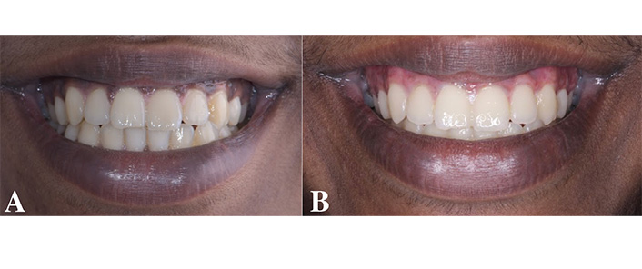
The perceptive smile aesthetics were evaluated using frontal smiling photographs. Photographs of each patient were taken before and after treatment at a consistent distance of 90 cm within the aesthetics department, utilizing ambient lighting conditions. The social smile deemed the most repeatable was documented for all patients [14]. The curriculum instructs all residents to document the social smile. The frontal smiling photos from the pre- and post-depigmentation stages were later imported in jpeg format into Adobe Photoshop (Adobe Photoshop CC version 14.0; Adobe Systems, San Jose, California). The photos were standardized to the same size (3 × 5 ratio) by cropping only the smile (Figure 1).

Standardized smile images. (A) pre-depigmentation; (B) post-depigmentation frontal smile images which were standardized and cropped
The photographs were randomly assigned numbers and included in two Google Forms for assessment by a panel of raters. The pre- and post-treatment photos were randomized to ensure that the evaluators remained unaware of the study’s objective. Two Google Forms were established to ensure that the pre- and post-depigmentation photos are not contained within the same form. The panel consisted of 40 adult laypersons with the minimum educational qualification of high school completion. Raters of both genders were randomly selected from the adult patients (above 20 years of age) visiting the hospital for treatment. All the raters were of Dravidian ethnicity. Written consent was obtained from the laypeople to participate in the study. Once the raters agreed to participate in the study, the Google Form URLs were disseminated to the raters via email and WhatsApp. All raters were calibrated according to the methodology outlined in research by Negreiros et al. [15]. Sample photographs displayed on the initial page of the Google link were utilized for inter-rater calibration of scores. The attractiveness of the smile was evaluated by assigning a score on a numeric scale from 1 to 10 on a 10-point Likert scale. The scores for every photograph varied from 1 to 10, with 1 representing the least appealing and 10 denoting the most attractive smile. The two Google Forms were sent out to the raters after a two-week interval between them. The forms were resent to the raters for reevaluation using the same approach as initially employed after two months. The mean scores were taken for statistical analysis.
The gingival pigmentation was assessed pre- and post-depigmentation surgery using the oral pigmentation index (OPI) [16]. The OPI scores were assessed using frontal intraoral images. An additional intraoral image exclusive of the image used in the study was used for assessment of inter-rater reliability. Based on the OPI ratings, both pre- and post-treatment photographs were scored as follows: score 0: absence of clinical pigmentation (pink gingiva); score 1: mild clinical pigmentation (light brown hue); score 2: moderate clinical pigmentation (medium brown or a combination of pink and brown); score 3: pronounced clinical pigmentation (deep brown or bluish-black hue). The pigmentation was assessed by three periodontists with more than five years of clinical and academic expertise, and the final scores were recorded based on consensus from the experts for all the samples pre- and post-treatment.
All scores were recorded in a Microsoft Excel spreadsheet. The Shapiro-Wilk test and Kolmogorov-Smirnov test confirmed the normality of the data. The Wilcoxon signed-rank test was employed to compare pre-treatment and post-treatment aesthetic and pigmentation scores. The Spearman correlation was used to detect correlation between gingival pigmentation scores and smile aesthetic scores. Inter-rater reliability for the aesthetic scores and OPI score was evaluated using the intraclass correlation coefficient (ICC). A significance criterion of p ≤ 0.05 was established.
The current investigation was carried out over a duration of six months. Fifteen pre-treatment and fifteen post-treatment photos were evaluated by laypersons. Out of 40 raters, only 30 completed all the questions, resulting in the analysis of 30 scores for statistical significance. A total of 10 raters did not respond to the forms even after reminder messages. All raters were adults, with a mean age of 30.3 years. The rater panel consisted of 14 females and 16 males.
The OPI scores were not normally distributed based on the normality analysis and hence a Wilcoxon signed-rank test was used for analysis. The mean OPI scores prior to depigmentation were 2.67 ± 0.49, however, post-depigmentation, the scores drastically decreased to a mean of 0.33 ± 0.5. The decrease in scores before and after depigmentation was statistically significant (p < 0.001). The smile aesthetic scores were also compared using the Wilcoxon signed-rank test. The average smile aesthetic ratings for the pre-depigmentation group were 6.72 ± 0.32, but the scores for the post-depigmentation group were higher with a mean score of 7.91 ± 0.18. There was a statistically significant difference in smile aesthetics scores between the pre- and post-depigmentation groups (p = 0.001) (Table 1).
Pre- and post-depigmentation OPI scores and smile aesthetic scores were compared using Wilcoxon signed-rank test
| Parameter | Pre-depigmentation | Post-depigmentation | p value | ||
|---|---|---|---|---|---|
| Mean and SD | Confidence interval | Mean and SD | Confidence interval | ||
| OPI scores | 2.67 ± 0.49 | 2.4–2.9 | 0.33 ± 0.5 | 0.0631–0.6036 | < 0.001** |
| Smile aesthetic scores | 6.72 ± 0.32 | 6.5–6.9 | 7.91 ± 0.18 | 7.8–8.0 | 0.001** |
OPI: oral pigmentation index; SD: standard deviation; ** indicates statistical significance
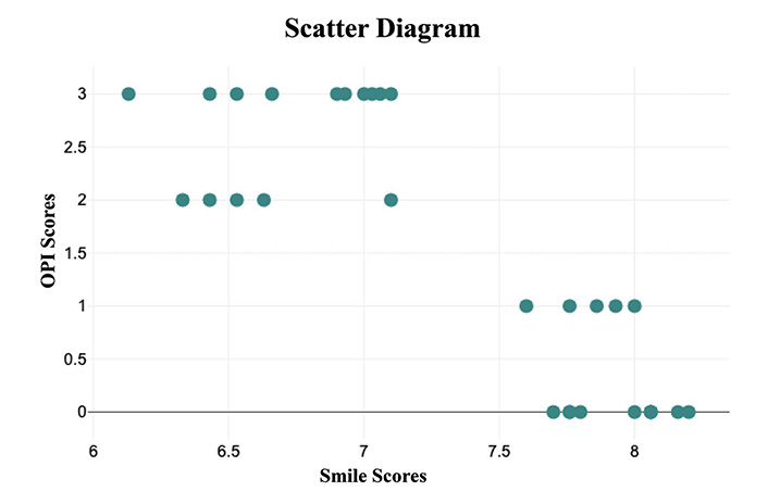
The Spearman correlation test revealed a negative correlation (r = –0.76) between smile aesthetics ratings and OPI scores, which was statistically significant (p < 0.001) (Figure 2). Good agreement (ICC = 0.756) was seen between the first rating and the rating of smile attractiveness performed two months later, among the raters. An ICC score of 0.923 was seen regarding the OPI scores amongst the three raters indicating excellent inter-rater reliability.

The result of the Spearman correlation as indicated in the scatter plot showed that there was a very high, negative correlation between smile aesthetic scores and oral pigmentation index (OPI) scores with r = –0.76
Gingival hyperpigmentation is an aesthetic concern for many patients, prompting several of them to seek consultations with periodontists and cosmetic dentists for improving their smiles. This is genetically determined in certain populations and is also called physiological or racial pigmentation and may correlate with the overall facial complexion [6]. This observational research assesses laypeople’s views on gingival pigmentation in South India. The study included retrospective photographs of patients’ smiles who received gingival depigmentation following orthodontic smile restoration. This was executed to eliminate additional variables that affect the aesthetic ratings of smiles. All included patients exhibited optimal dental relationships in accordance with Andrew’s six keys of occlusion and were treated under the supervision of orthodontists with over 10 years of expertise. The systematic review by Coppola et al. [17] indicates that orthodontic smile correction has a moderate and favorable impact on perceived smile aesthetics. Consequently, this sample was employed to evaluate the real influence of gingival pigmentation on smile aesthetic ratings.
The present study demonstrated a significant difference in the perceived aesthetic evaluations of smiles prior to and following depigmentation and revealed an inverse relationship between smile aesthetic perceptions and the degree of oral pigmentation. A previous study by Batra et al. [10] evaluated the effect of altered gingival aesthetics on laypersons’ judgments of smile aesthetics. The study demonstrated that laypersons were especially observant of changes in the color of the gingiva. The alterations in gingival color due to pigmentation obtained low ratings and were thus considered significantly unaesthetic, like the results of the present study. Alomari et al. [3] found that non-experts more readily recognized changes in gingival color due to pigmentation as unaesthetic, compared to other gingival characteristics like gingival contour and zenith. Sharma et al. [12] validated similar results in an additional investigation. However, in contrast to the present study, all previous research used an image that was specifically deemed attractive and digitally altered the smiling attributes for assessment. In a separate study by Siraj et al. [18], the vermilion color and gingival pigmentation deemed most aesthetic were evaluated. The research revealed that people deemed pigmented lips and gingiva unaesthetic, which is in line with the current study’s findings about gingival pigmentation. Separate research by Ashok et al. [19] and Prashaanthi and Kaarthikeyan [20] also showed that young people generally perceived pigmented gingiva as unappealing, consistent with the current study’s findings, and were willing to undergo treatment.
The present study demonstrates that all patients who had depigmentation exhibited moderate to severe pre-treatment gingival pigmentation scores, with a mean OPI score of 2.67 ± 0.49, which considerably decreased following the procedure. Due to the retrospective acquisition of data, the depigmentation approach was not standardized throughout the evaluated samples, with most patients undergoing laser-assisted gingival depigmentation. According to the systematic review by Lin et al. [21], cryosurgery, electrosurgery, and laser surgery are the most effective treatment techniques for gingival pigmentation. All the cases included in the study were treated using laser depigmentation. The current study employed the OPI to evaluate gingival pigmentation due to its wide adoption and its simplicity and convenience of usage.
The current study elucidates a significant gingival characteristic that may adversely affect the impression of smile aesthetics among South Indian laypeople specifically. The strength of the current study is in the utilization of real-world scenario based pre- and post-depigmentation photographs, as opposed to digitally manipulated photos employed in previous studies for perceptual analysis. All post-depigmentation photos were documented within a period of two months. This was conducted with the consideration that gingival re-pigmentation may transpire within one year of therapy [10]. Nonetheless, the study had several limitations due to the retrospective data acquisition strategy since patient parameters could not be standardized. The sample size also was limited. The findings may be further verified with a larger, ethnically diverse population. Future studies with adequate sample sizes can be done to generalize the findings of the present study. The present study used only one index, the OPI, to evaluate the gingival pigmentation. However, researchers could consider using more comprehensive indices such as the gingival pigmentation and pigmented lesions index. Additionally, the study results may be biased due to cultural and ethnic factors. Further studies must be undertaken with raters of varying cultural and ethnic backgrounds. The significance of pink aesthetics in smile designs is increasing. Hence, additional research examining the impact of various gingival factors on smile aesthetics is necessary. Specifically, characteristics like gingival pigmentation, a natural variation, do not require any intervention. The psychological causes and implications of the patient perception must be investigated in detail. Also, future studies must be conducted prospectively to understand the psychological causes and implications of the patient’s own perception in detail. Nonetheless, a patient’s impression may differ from that of the professional, and addressing this discrepancy can enhance overall treatment outcomes and patient satisfaction.
In conclusion, within the scope of the present study, laypersons perceived gingival pigmentation unfavorably, and smile aesthetic ratings were inversely related to the extent of hyperpigmentation. Depigmentation procedures may be suggested in such patients for a satisfactory smile correction, particularly in patients with excessive gingival show during a smile.
ICC: intraclass correlation coefficient
OPI: oral pigmentation index
PR: Data curation, Formal analysis, Investigation, Methodology, Writing—original draft, Resources. SN: Conceptualization, Project administration, Supervision, Validation, Visualization, Writing—original draft, Writing—review & editing. RB: Project administration, Supervision, Validation, Visualization, Writing—review & editing.
The authors declare that they have no conflicts of interest.
This study was approved by the Saveetha Dental College Institutional Ethical Committee (SRB/SDC/UG-2028/24/PEDO/309) and was conducted in accordance with the ethical principles outlined in the Declaration of Helsinki.
Informed consent to participate in the study was obtained from all participants.
Informed consent to publication was obtained from relevant participants.
The data that support the findings of this study are not publicly available due to privacy concerns and the confidential nature of participant information. To protect the privacy of the individuals involved, the data will remain confidential and will not be shared publicly.
Not applicable.
© The Author(s) 2025.
Open Exploration maintains a neutral stance on jurisdictional claims in published institutional affiliations and maps. All opinions expressed in this article are the personal views of the author(s) and do not represent the stance of the editorial team or the publisher.
Copyright: © The Author(s) 2025. This is an Open Access article licensed under a Creative Commons Attribution 4.0 International License (https://creativecommons.org/licenses/by/4.0/), which permits unrestricted use, sharing, adaptation, distribution and reproduction in any medium or format, for any purpose, even commercially, as long as you give appropriate credit to the original author(s) and the source, provide a link to the Creative Commons license, and indicate if changes were made.
View: 2508
Download: 37
Times Cited: 0
